Optimization of Triazine Nitriles as Rhodesain Inhibitors: Structure-Activity Relationships, Bioisosteric Imidazopyridine Nitriles, and X-Ray Crystal Structure Analysis with Human Cathepsin L
Ehmke, V., Winkler, E., Banner, D.W., Haap, W., Schweizer, W.B., Rottmann, M., Kaiser, M., Freymond, C., Schirmeister, T., Diederich, F.(2013) ChemMedChem 8: 967
- PubMed: 23658062
- DOI: https://doi.org/10.1002/cmdc.201300112
- Primary Citation of Related Structures:
4AXL, 4AXM - PubMed Abstract:
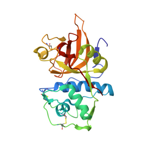
The cysteine protease rhodesain of Trypanosoma brucei parasites causing African sleeping sickness has emerged as a target for the development of new drug candidates. Based on a triazine nitrile moiety as electrophilic headgroup, optimization studies on the substituents for the S1, S2, and S3 pockets of the enzyme were performed using structure-based design and resulted in inhibitors with inhibition constants in the single-digit nanomolar range. Comprehensive structure-activity relationships clarified the binding preferences of the individual pockets of the active site. The S1 pocket tolerates various substituents with a preference for flexible and basic side chains. Variation of the S2 substituent led to high-affinity ligands with inhibition constants down to 2 nM for compounds bearing cyclohexyl substituents. Systematic investigations on the S3 pocket revealed its potential to achieve high activities with aromatic vectors that undergo stacking interactions with the planar peptide backbone forming part of the pocket. X-ray crystal structure analysis with the structurally related enzyme human cathepsin L confirmed the binding mode of the triazine ligand series as proposed by molecular modeling. Sub-micromolar inhibition of the proliferation of cultured parasites was achieved for ligands decorated with the best substituents identified through the optimization cycles. In cell-based assays, the introduction of a basic side chain on the inhibitors resulted in a 35-fold increase in antitrypanosomal activity. Finally, bioisosteric imidazopyridine nitriles were studied in order to prevent off-target effects with unselective nucleophiles by decreasing the inherent electrophilicity of the triazine nitrile headgroup. Using this ligand, the stabilization by intramolecular hydrogen bonding of the thioimidate intermediate, formed upon attack of the catalytic cysteine residue, compensates for the lower reactivity of the headgroup. The imidazopyridine nitrile ligand showed excellent stability toward the thiol nucleophile glutathione in a quantitative in vitro assay and fourfold lower cytotoxicity than the parent triazine nitrile.
Organizational Affiliation:
Laboratorium für Organische Chemie, ETH Zürich, Zürich, Switzerland.


