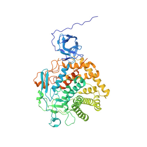
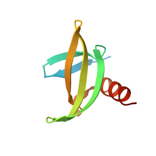
4CJ1

Crystal structure of CelD in complex with affitin H3


Experimental Data Snapshot

  • Method: X-RAY DIFFRACTION
  • Resolution: 1.63 Å
  • R-Value Free: 0.144 
  • R-Value Work: 0.106 
  • R-Value Observed: 0.108 

Starting Model: experimental
View more details

wwPDB Validation   3D Report Full Report


This is version 1.3 of the entry. See complete history


Literature

Potent and Specific Inhibition of Glycosidases by Small Artificial Binding Proteins (Affitins)

Correa, A.Pacheco, S.Mechaly, A.E.Obal, G.Behar, G.Mouratou, B.Oppezzo, P.Alzari, P.M.Pecorari, F.

(2014) PLoS One 9: 97438

  • DOI: https://doi.org/10.1371/journal.pone.0097438
  • Primary Citation of Related Structures:  
    4CJ0, 4CJ1, 4CJ2

  • PubMed Abstract: 

    Glycosidases are associated with various human diseases. The development of efficient and specific inhibitors may provide powerful tools to modulate their activity. However, achieving high selectivity is a major challenge given that glycosidases with different functions can have similar enzymatic mechanisms and active-site architectures. As an alternative approach to small-chemical compounds, proteinaceous inhibitors might provide a better specificity by involving a larger surface area of interaction. We report here the design and characterization of proteinaceous inhibitors that specifically target endoglycosidases representative of the two major mechanistic classes; retaining and inverting glycosidases. These inhibitors consist of artificial affinity proteins, Affitins, selected against the thermophilic CelD from Clostridium thermocellum and lysozyme from hen egg. They were obtained from libraries of Sac7d variants, which involve either the randomization of a surface or the randomization of a surface and an artificially-extended loop. Glycosidase binders exhibited affinities in the nanomolar range with no cross-recognition, with efficient inhibition of lysozyme (Ki = 45 nM) and CelD (Ki = 95 and 111 nM), high expression yields in Escherichia coli, solubility, and thermal stabilities up to 81.1°C. The crystal structures of glycosidase-Affitin complexes validate our library designs. We observed that Affitins prevented substrate access by two modes of binding; covering or penetrating the catalytic site via the extended loop. In addition, Affitins formed salt-bridges with residues essential for enzymatic activity. These results lead us to propose the use of Affitins as versatile selective glycosidase inhibitors and, potentially, as enzymatic inhibitors in general.


  • Organizational Affiliation

    Institut Pasteur de Montevideo, Recombinant Protein Unit, Montevideo, Uruguay; Institut Pasteur, Unité de Microbiologie Structurale, CNRS UMR 3528, Paris, France.


Macromolecules
Find similar proteins by:  (by identity cutoff)  |  3D Structure
Entity ID: 1
MoleculeChains Sequence LengthOrganismDetailsImage
ENDOGLUCANASE D625Acetivibrio thermocellusMutation(s): 0 
EC: 3.2.1.4
UniProt
Find proteins for P0C2S4 (Acetivibrio thermocellus)
Explore P0C2S4 
Go to UniProtKB:  P0C2S4
Entity Groups  
Sequence Clusters30% Identity50% Identity70% Identity90% Identity95% Identity100% Identity
UniProt GroupP0C2S4
Sequence Annotations
Expand
  • Reference Sequence
Find similar proteins by:  (by identity cutoff)  |  3D Structure
Entity ID: 2
MoleculeChains Sequence LengthOrganismDetailsImage
H3 AFFITIN81synthetic constructMutation(s): 0 
UniProt
Find proteins for P13123 (Sulfolobus acidocaldarius (strain ATCC 33909 / DSM 639 / JCM 8929 / NBRC 15157 / NCIMB 11770))
Explore P13123 
Go to UniProtKB:  P13123
Entity Groups  
Sequence Clusters30% Identity50% Identity70% Identity90% Identity95% Identity100% Identity
UniProt GroupP13123
Sequence Annotations
Expand
  • Reference Sequence
Small Molecules
Ligands 3 Unique
IDChains Name / Formula / InChI Key2D Diagram3D Interactions
GOL
Query on GOL

Download Ideal Coordinates CCD File 
G [auth A],
H [auth A],
I [auth A],
J [auth A]
GLYCEROL
C3 H8 O3
PEDCQBHIVMGVHV-UHFFFAOYSA-N
ZN
Query on ZN

Download Ideal Coordinates CCD File 
C [auth A]ZINC ION
Zn
PTFCDOFLOPIGGS-UHFFFAOYSA-N
CA
Query on CA

Download Ideal Coordinates CCD File 
D [auth A],
E [auth A],
F [auth A]
CALCIUM ION
Ca
BHPQYMZQTOCNFJ-UHFFFAOYSA-N
Experimental Data & Validation

Experimental Data

  • Method: X-RAY DIFFRACTION
  • Resolution: 1.63 Å
  • R-Value Free: 0.144 
  • R-Value Work: 0.106 
  • R-Value Observed: 0.108 
  • Space Group: P 21 21 21
Unit Cell:
Length ( Å )Angle ( ˚ )
a = 74.422α = 90
b = 97.736β = 90
c = 106.58γ = 90
Software Package:
Software NamePurpose
REFMACrefinement
XDSdata reduction
Aimlessdata scaling
PHASERphasing

Structure Validation

View Full Validation Report


Entry History 

Deposition Data

Revision History  (Full details and data files)

  • Version 1.0: 2014-05-21
    Type: Initial release
  • Version 1.1: 2014-05-28
    Changes: Database references
  • Version 1.2: 2018-05-16
    Changes: Data collection
  • Version 1.3: 2023-12-20
    Changes: Data collection, Database references, Derived calculations, Other, Refinement description