4CS0

Direct visualisation of strain-induced protein post-translational modification


Experimental Data Snapshot

  • Method: X-RAY DIFFRACTION
  • Resolution: 2.10 Å
  • R-Value Free: 0.237 
  • R-Value Work: 0.172 
  • R-Value Observed: 0.175 

Starting Model: experimental
View more details

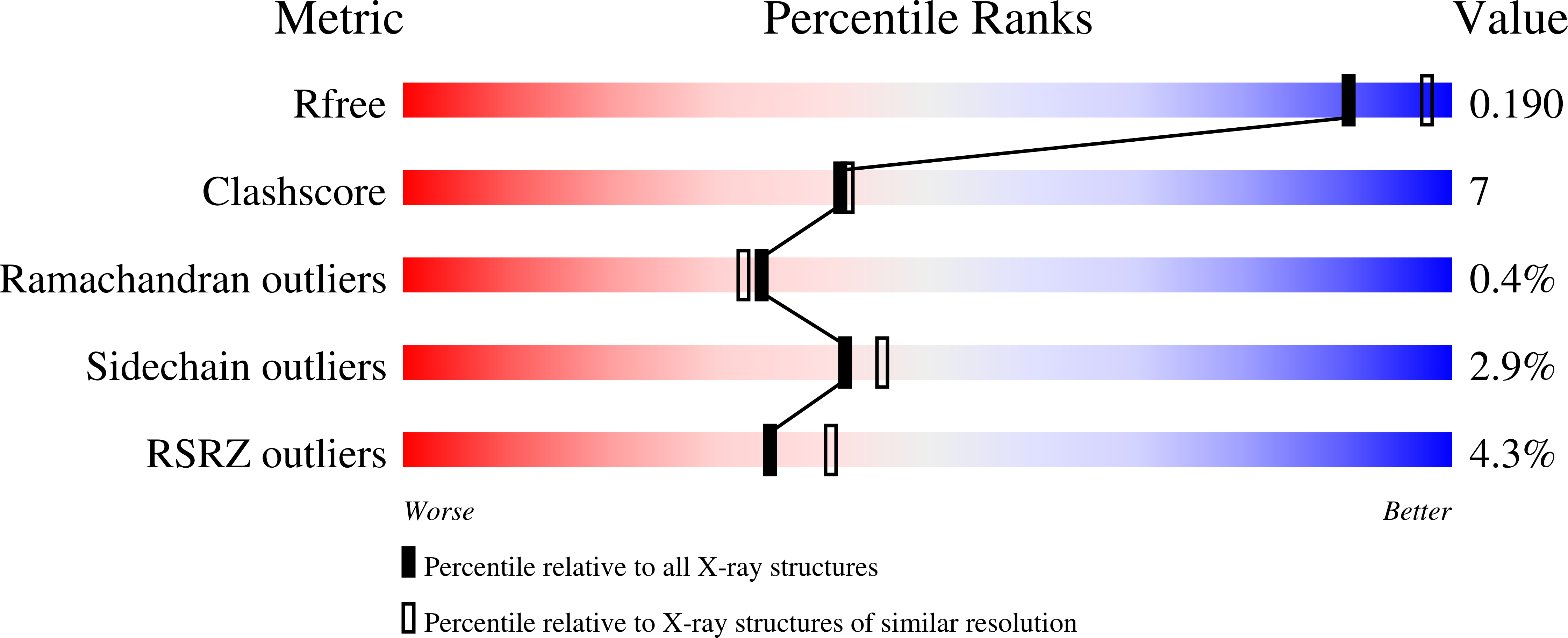
wwPDB Validation   3D Report Full Report


Ligand Structure Quality Assessment 


This is version 1.4 of the entry. See complete history


Literature

The Structure of the Pand/Panz Protein Complex Reveals Negative Feedback Regulation of Pantothenate Biosynthesis by Coenzyme A.

Monteiro, D.C.Patel, V.Bartlett, C.P.Nozaki, S.Grant, T.D.Gowdy, J.A.Thompson, G.S.Kalverda, A.P.Snell, E.H.Niki, H.Pearson, A.R.Webb, M.E.

(2015) Chem Biol 22: 492

  • DOI: https://doi.org/10.1016/j.chembiol.2015.03.017
  • Primary Citation of Related Structures:  
    4CRZ, 4CS0

  • PubMed Abstract: 

    Coenzyme A (CoA) is an ubiquitous and essential cofactor, synthesized from the precursor pantothenate. Vitamin biosynthetic pathways are normally tightly regulated, including the pathway from pantothenate to CoA. However, no regulation of pantothenate biosynthesis has been identified. We have recently described an additional component in the pantothenate biosynthetic pathway, PanZ, which promotes the activation of the zymogen, PanD, to form aspartate α-decarboxylase (ADC) in a CoA-dependent manner. Here we report the structure of PanZ in complex with PanD, which reveals the structural basis for the CoA dependence of this interaction and activation. In addition, we show that PanZ acts as a CoA-dependent inhibitor of ADC catalysis. This inhibitory effect can effectively regulate the biosynthetic pathway to pantothenate, and thereby also regulate CoA biosynthesis. This represents a previously unobserved mode of metabolic regulation whereby a cofactor-utilizing protein negatively regulates the biosynthesis of the same cofactor.


  • Organizational Affiliation

    Astbury Centre for Structural Molecular Biology, University of Leeds, Leeds LS2 9JT, UK; School of Chemistry, University of Leeds, Leeds LS2 9JT, UK; Faculty of Biological Sciences, University of Leeds, Leeds LS2 9JT, UK.


Macromolecules
Find similar proteins by:  (by identity cutoff)  |  3D Structure
Entity ID: 1
MoleculeChains Sequence LengthOrganismDetailsImage
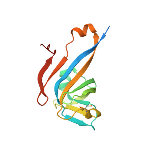
ASPARTATE 1-DECARBOXYLASE143Escherichia coli K-12Mutation(s): 1 
EC: 4.1.1.11
UniProt
Find proteins for P0A790 (Escherichia coli (strain K12))
Explore P0A790 
Go to UniProtKB:  P0A790
Entity Groups  
Sequence Clusters30% Identity50% Identity70% Identity90% Identity95% Identity100% Identity
UniProt GroupP0A790
Sequence Annotations
Expand
  • Reference Sequence
Find similar proteins by:  (by identity cutoff)  |  3D Structure
Entity ID: 2
MoleculeChains Sequence LengthOrganismDetailsImage
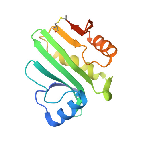
PANZ137Escherichia coli K-12Mutation(s): 0 
UniProt
Find proteins for P37613 (Escherichia coli (strain K12))
Explore P37613 
Go to UniProtKB:  P37613
Entity Groups  
Sequence Clusters30% Identity50% Identity70% Identity90% Identity95% Identity100% Identity
UniProt GroupP37613
Sequence Annotations
Expand
  • Reference Sequence
Experimental Data & Validation

Experimental Data

  • Method: X-RAY DIFFRACTION
  • Resolution: 2.10 Å
  • R-Value Free: 0.237 
  • R-Value Work: 0.172 
  • R-Value Observed: 0.175 
  • Space Group: I 4
Unit Cell:
Length ( Å )Angle ( ˚ )
a = 86.27α = 90
b = 86.27β = 90
c = 80.81γ = 90
Software Package:
Software NamePurpose
REFMACrefinement
iMOSFLMdata reduction
Aimlessdata scaling
MOLREPphasing

Structure Validation

View Full Validation Report


Ligand Structure Quality Assessment 


Entry History 

Deposition Data

Revision History  (Full details and data files)

  • Version 1.0: 2015-03-25
    Type: Initial release
  • Version 1.1: 2015-05-06
    Changes: Database references
  • Version 1.2: 2017-06-28
    Changes: Data collection
  • Version 1.3: 2023-12-20
    Changes: Data collection, Database references, Derived calculations, Other, Refinement description
  • Version 1.4: 2024-11-20
    Changes: Structure summary