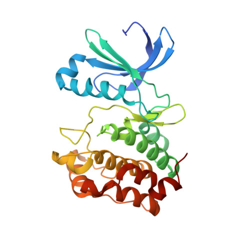
4DHF

Structure of Aurora A mutant bound to Biogenidec cpd 15


Experimental Data Snapshot

  • Method: X-RAY DIFFRACTION
  • Resolution: 2.80 Å
  • R-Value Free: 0.299 
  • R-Value Work: 0.206 
  • R-Value Observed: 0.210 

wwPDB Validation   3D Report Full Report


Ligand Structure Quality Assessment 


This is version 1.1 of the entry. See complete history


Literature

Structure-based design of 2,6,7-trisubstituted-7H-pyrrolo[2,3-d]pyrimidines as Aurora kinases inhibitors.

Le Brazidec, J.Y.Pasis, A.Tam, B.Boykin, C.Wang, D.Marcotte, D.J.Claassen, G.Chong, J.H.Chao, J.Fan, J.Nguyen, K.Silvian, L.Ling, L.Zhang, L.Choi, M.Teng, M.Pathan, N.Zhao, S.Li, T.Taveras, A.

(2012) Bioorg Med Chem Lett 22: 4033-4037

  • DOI: https://doi.org/10.1016/j.bmcl.2012.04.085
  • Primary Citation of Related Structures:  
    4DHF

  • PubMed Abstract: 

    This Letter reports the optimization of a pyrrolopyrimidine series as dual inhibitors of Aurora A/B kinases. This series derived from a pyrazolopyrimidine series previously reported as inhibitors of aurora kinases and CDKs. In an effort to improve the selectivity of this chemotype, we switched to the pyrrolopyrimidine core which allowed functionalization on C-2. In addition, the modeling rationale was based on superimposing the structures of Aurora-A kinase and CDK2 which revealed enough differences leading to a path for selectivity improvement. The synthesis of the new series of pyrrolopyrimidine analogs relied on the development of a different route for the two key intermediates 7 and 19 which led to analogs with both tunable activity against CDK1 and maintained cell potency.


  • Organizational Affiliation

    Biogen Idec, 5200 Research Place, San Diego, CA 92122, USA. [email protected]


Macromolecules
Find similar proteins by:  (by identity cutoff)  |  3D Structure
Entity ID: 1
MoleculeChains Sequence LengthOrganismDetailsImage
Aurora kinase A
A, B
271Homo sapiensMutation(s): 2 
Gene Names: 
AIKAIRK1ARK1AURAAURKAAuroraAAYK1BTAKIAK1STK15...
AIKAIRK1ARK1AURAAURKAAuroraAAYK1BTAKIAK1STK15STK6

EC: 2.7.11.1
UniProt & NIH Common Fund Data Resources
Find proteins for O14965 (Homo sapiens)
Explore O14965 
Go to UniProtKB:  O14965
PHAROS:  O14965
GTEx:  ENSG00000087586 
Entity Groups  
Sequence Clusters30% Identity50% Identity70% Identity90% Identity95% Identity100% Identity
UniProt GroupO14965
Sequence Annotations
Expand
  • Reference Sequence
Small Molecules
Ligands 3 Unique
IDChains Name / Formula / InChI Key2D Diagram3D Interactions
0K6
Query on 0K6

Download Ideal Coordinates CCD File 
C [auth A],
F [auth B]
7-cyclopentyl-2-({1-methyl-5-[(4-methylpiperazin-1-yl)carbonyl]-1H-pyrrol-3-yl}amino)-7H-pyrrolo[2,3-d]pyrimidine-6-carboxamide
C23 H30 N8 O2
CXCXNZWXDOVZBA-UHFFFAOYSA-N
PO4
Query on PO4

Download Ideal Coordinates CCD File 
D [auth A],
G [auth B]
PHOSPHATE ION
O4 P
NBIIXXVUZAFLBC-UHFFFAOYSA-K
MG
Query on MG

Download Ideal Coordinates CCD File 
E [auth A],
H [auth B]
MAGNESIUM ION
Mg
JLVVSXFLKOJNIY-UHFFFAOYSA-N
Binding Affinity Annotations 
IDSourceBinding Affinity
0K6 PDBBind:  4DHF IC50: 28 (nM) from 1 assay(s)
Experimental Data & Validation

Experimental Data

  • Method: X-RAY DIFFRACTION
  • Resolution: 2.80 Å
  • R-Value Free: 0.299 
  • R-Value Work: 0.206 
  • R-Value Observed: 0.210 
  • Space Group: P 61
Unit Cell:
Length ( Å )Angle ( ˚ )
a = 81.59α = 90
b = 81.59β = 90
c = 166.065γ = 120
Software Package:
Software NamePurpose
MOLREPphasing
REFMACrefinement
HKL-2000data reduction
HKL-2000data scaling

Structure Validation

View Full Validation Report


Ligand Structure Quality Assessment 


Entry History 

Deposition Data

Revision History  (Full details and data files)

  • Version 1.0: 2012-07-18
    Type: Initial release
  • Version 1.1: 2024-02-28
    Changes: Data collection, Database references, Derived calculations