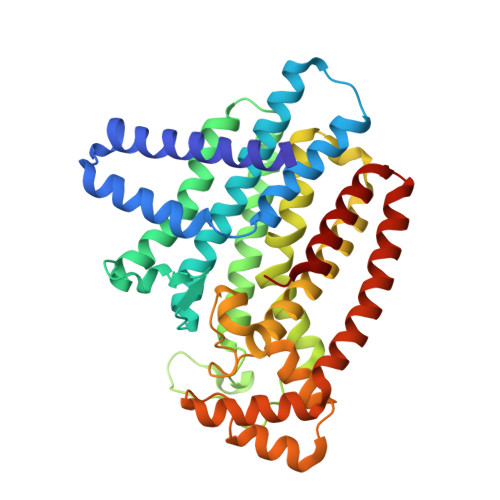
4DXJ

Crystal structure of Trypanosome cruzi farnesyl diphosphate synthase in complex with [2-(n-propylamino)ethane-1,1-diyl]bisphosphonic acid and Mg2+


Experimental Data Snapshot

  • Method: X-RAY DIFFRACTION
  • Resolution: 2.35 Å
  • R-Value Free: 0.285 
  • R-Value Work: 0.220 
  • R-Value Observed: 0.223 

Starting Model: experimental
View more details

wwPDB Validation   3D Report Full Report


Ligand Structure Quality Assessment 


This is version 1.2 of the entry. See complete history


Literature

Design, synthesis, calorimetry, and crystallographic analysis of 2-alkylaminoethyl-1,1-bisphosphonates as inhibitors of Trypanosoma cruzi farnesyl diphosphate synthase.

Aripirala, S.Szajnman, S.H.Jakoncic, J.Rodriguez, J.B.Docampo, R.Gabelli, S.B.Amzel, L.M.

(2012) J Med Chem 55: 6445-6454

  • DOI: https://doi.org/10.1021/jm300425y
  • Primary Citation of Related Structures:  
    4DWB, 4DWG, 4DXJ, 4DZW, 4E1E

  • PubMed Abstract: 

    Linear 2-alkylaminoethyl-1,1-bisphosphonates are effective agents against proliferation of Trypanosoma cruzi , the etiologic agent of American trypanosomiasis (Chagas disease), exhibiting IC(50) values in the nanomolar range against the parasites. This activity is associated with inhibition at the low nanomolar level of the T. cruzi farnesyl diphosphate synthase (TcFPPS). X-ray structures and thermodynamic data of the complexes TcFPPS with five compounds of this family show that the inhibitors bind to the allylic site of the enzyme, with their alkyl chain occupying the cavity that binds the isoprenoid chain of the substrate. The compounds bind to TcFPPS with unfavorable enthalpy compensated by a favorable entropy that results from a delicate balance between two opposing effects: the loss of conformational entropy due to freezing of single bond rotations and the favorable burial of the hydrophobic alkyl chains. The data suggest that introduction of strategically placed double bonds and methyl branches should increase affinity substantially.


  • Organizational Affiliation

    Department of Biophysics and Biophysical Chemistry, Johns Hopkins University School of Medicine, Baltimore, MD 21205, USA.


Macromolecules
Find similar proteins by:  (by identity cutoff)  |  3D Structure
Entity ID: 1
MoleculeChains Sequence LengthOrganismDetailsImage
Farnesyl pyrophosphate synthase
A, B, C
362Trypanosoma cruziMutation(s): 0 
Gene Names: FPPS
EC: 2.5.1.10
UniProt
Find proteins for Q95WL3 (Trypanosoma cruzi)
Explore Q95WL3 
Go to UniProtKB:  Q95WL3
Entity Groups  
Sequence Clusters30% Identity50% Identity70% Identity90% Identity95% Identity100% Identity
UniProt GroupQ95WL3
Sequence Annotations
Expand
  • Reference Sequence
Small Molecules
Ligands 7 Unique
IDChains Name / Formula / InChI Key2D Diagram3D Interactions
0M9
Query on 0M9

Download Ideal Coordinates CCD File 
G [auth A],
N [auth B],
W [auth C]
[2-(propylamino)ethane-1,1-diyl]bis(phosphonic acid)
C5 H15 N O6 P2
YFFJUBPLIKXHAO-UHFFFAOYSA-N
IPE
Query on IPE

Download Ideal Coordinates CCD File 
H [auth A],
O [auth B],
X [auth C]
3-METHYLBUT-3-ENYL TRIHYDROGEN DIPHOSPHATE
C5 H12 O7 P2
NUHSROFQTUXZQQ-UHFFFAOYSA-N
PGE
Query on PGE

Download Ideal Coordinates CCD File 
S [auth B]TRIETHYLENE GLYCOL
C6 H14 O4
ZIBGPFATKBEMQZ-UHFFFAOYSA-N
PEG
Query on PEG

Download Ideal Coordinates CCD File 
AA [auth C],
I [auth A],
P [auth B],
Q [auth B],
Z [auth C]
DI(HYDROXYETHYL)ETHER
C4 H10 O3
MTHSVFCYNBDYFN-UHFFFAOYSA-N
SO4
Query on SO4

Download Ideal Coordinates CCD File 
J [auth A],
R [auth B]
SULFATE ION
O4 S
QAOWNCQODCNURD-UHFFFAOYSA-L
ACT
Query on ACT

Download Ideal Coordinates CCD File 
Y [auth C]ACETATE ION
C2 H3 O2
QTBSBXVTEAMEQO-UHFFFAOYSA-M
MG
Query on MG

Download Ideal Coordinates CCD File 
D [auth A]
E [auth A]
F [auth A]
K [auth B]
L [auth B]
D [auth A],
E [auth A],
F [auth A],
K [auth B],
L [auth B],
M [auth B],
T [auth C],
U [auth C],
V [auth C]
MAGNESIUM ION
Mg
JLVVSXFLKOJNIY-UHFFFAOYSA-N
Binding Affinity Annotations 
IDSourceBinding Affinity
0M9 PDBBind:  4DXJ IC50: 38 (nM) from 1 assay(s)
Experimental Data & Validation

Experimental Data

  • Method: X-RAY DIFFRACTION
  • Resolution: 2.35 Å
  • R-Value Free: 0.285 
  • R-Value Work: 0.220 
  • R-Value Observed: 0.223 
  • Space Group: P 61 2 2
Unit Cell:
Length ( Å )Angle ( ˚ )
a = 103.22α = 90
b = 103.22β = 90
c = 386.658γ = 120
Software Package:
Software NamePurpose
SCALEPACKdata scaling
REFMACrefinement
PDB_EXTRACTdata extraction
CBASSdata collection
DENZOdata reduction
REFMACphasing

Structure Validation

View Full Validation Report


Ligand Structure Quality Assessment 


Entry History 

Deposition Data

Revision History  (Full details and data files)

  • Version 1.0: 2013-01-16
    Type: Initial release
  • Version 1.1: 2013-04-17
    Changes: Non-polymer description
  • Version 1.2: 2023-09-13
    Changes: Data collection, Database references, Derived calculations, Refinement description