4EG1

Trypanosoma brucei methionyl-tRNA synthetase in complex with substrate Methionine


Experimental Data Snapshot

  • Method: X-RAY DIFFRACTION
  • Resolution: 2.90 Å
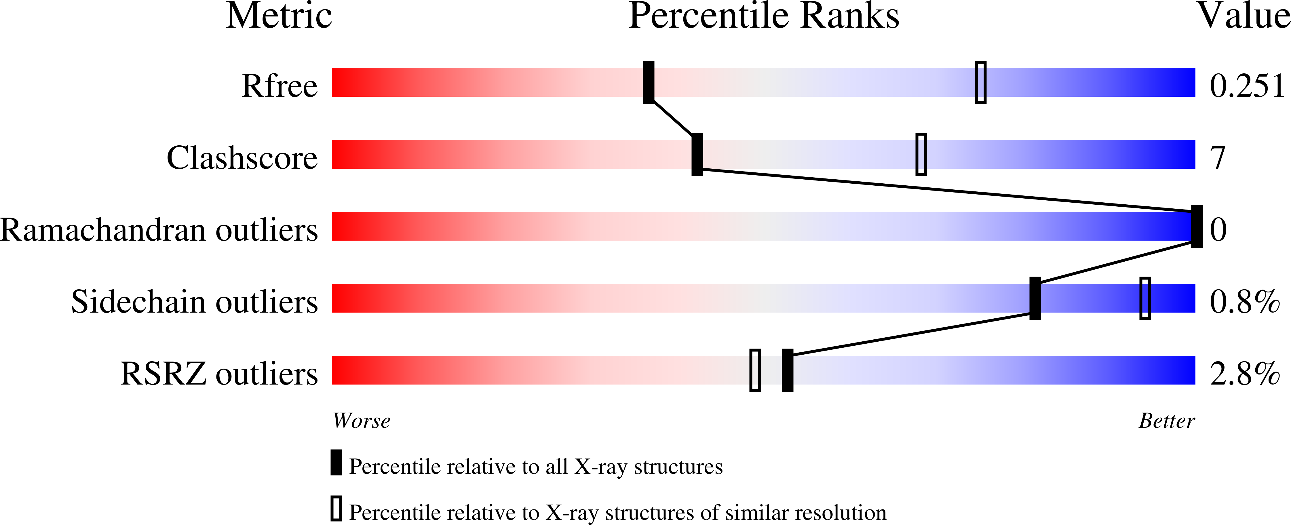
  • R-Value Free: 0.254 
  • R-Value Work: 0.209 
  • R-Value Observed: 0.211 

Starting Model: experimental
View more details

wwPDB Validation   3D Report Full Report


This is version 1.5 of the entry. See complete history


Literature

Distinct States of Methionyl-tRNA Synthetase Indicate Inhibitor Binding by Conformational Selection.

Koh, C.Y.Kim, J.E.Shibata, S.Ranade, R.M.Yu, M.Liu, J.Gillespie, J.R.Buckner, F.S.Verlinde, C.L.Fan, E.Hol, W.G.

(2012) Structure 20: 1681-1691

  • DOI: https://doi.org/10.1016/j.str.2012.07.011
  • Primary Citation of Related Structures:  
    4EG1, 4EG3, 4EG4, 4EG5, 4EG6, 4EG7, 4EG8, 4EGA

  • PubMed Abstract: 

    To guide development of new drugs targeting methionyl-tRNA synthetase (MetRS) for treatment of human African trypanosomiasis, crystal structure determinations of Trypanosoma brucei MetRS in complex with its substrate methionine and its intermediate product methionyl-adenylate were followed by those of the enzyme in complex with high-affinity aminoquinolone inhibitors via soaking experiments. Drastic changes in conformation of one of the two enzymes in the asymmetric unit allowed these inhibitors to occupy an enlarged methionine pocket and a new so-called auxiliary pocket. Interestingly, a small low-affinity compound caused the same conformational changes, removed the methionine without occupying the methionine pocket, and occupied the previously not existing auxiliary pocket. Analysis of these structures indicates that the binding of the inhibitors is the result of conformational selection, not induced fit.


  • Organizational Affiliation

    Department of Biochemistry, University of Washington, Seattle, WA 98195, USA.


Macromolecules
Find similar proteins by:  (by identity cutoff)  |  3D Structure
Entity ID: 1
MoleculeChains Sequence LengthOrganismDetailsImage
Methionyl-tRNA synthetase, putative
A, B
542Trypanosoma brucei brucei TREU927Mutation(s): 3 
Gene Names: Tb10.70.6470
EC: 6.1.1.10
UniProt
Find proteins for Q38C91 (Trypanosoma brucei brucei (strain 927/4 GUTat10.1))
Explore Q38C91 
Go to UniProtKB:  Q38C91
Entity Groups  
Sequence Clusters30% Identity50% Identity70% Identity90% Identity95% Identity100% Identity
UniProt GroupQ38C91
Sequence Annotations
Expand
  • Reference Sequence
Small Molecules
Ligands 2 Unique
IDChains Name / Formula / InChI Key2D Diagram3D Interactions
MET
Query on MET

Download Ideal Coordinates CCD File 
J [auth A],
P [auth B]
METHIONINE
C5 H11 N O2 S
FFEARJCKVFRZRR-BYPYZUCNSA-N
GOL
Query on GOL

Download Ideal Coordinates CCD File 
C [auth A]
D [auth A]
E [auth A]
F [auth A]
G [auth A]
C [auth A],
D [auth A],
E [auth A],
F [auth A],
G [auth A],
H [auth A],
I [auth A],
K [auth B],
L [auth B],
M [auth B],
N [auth B],
O [auth B]
GLYCEROL
C3 H8 O3
PEDCQBHIVMGVHV-UHFFFAOYSA-N
Modified Residues  1 Unique
IDChains TypeFormula2D DiagramParent
CAS
Query on CAS
A, B
L-PEPTIDE LINKINGC5 H12 As N O2 SCYS
Experimental Data & Validation

Experimental Data

  • Method: X-RAY DIFFRACTION
  • Resolution: 2.90 Å
  • R-Value Free: 0.254 
  • R-Value Work: 0.209 
  • R-Value Observed: 0.211 
  • Space Group: P 21 21 21
Unit Cell:
Length ( Å )Angle ( ˚ )
a = 85.037α = 90
b = 105.935β = 90
c = 207.18γ = 90
Software Package:
Software NamePurpose
DENZOdata reduction
SCALEPACKdata scaling
PHASERphasing
REFMACrefinement
PDB_EXTRACTdata extraction

Structure Validation

View Full Validation Report


Entry History 

Deposition Data

Revision History  (Full details and data files)

  • Version 1.0: 2012-09-12
    Type: Initial release
  • Version 1.1: 2012-10-17
    Changes: Other
  • Version 1.2: 2012-10-31
    Changes: Database references
  • Version 1.3: 2017-11-15
    Changes: Refinement description
  • Version 1.4: 2023-09-13
    Changes: Data collection, Database references, Derived calculations, Refinement description
  • Version 1.5: 2024-11-06
    Changes: Structure summary