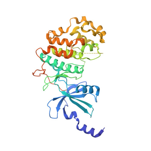
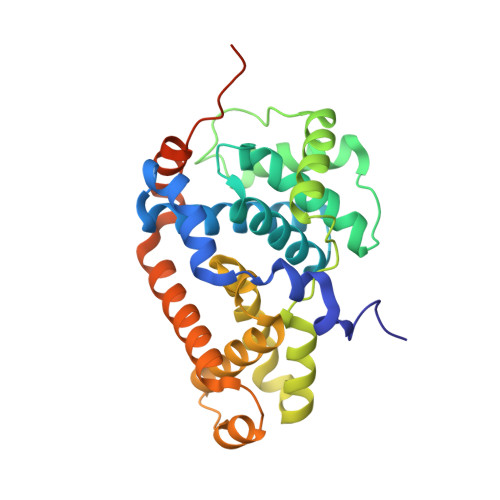
4F7J

Crystal structure of human CDK8/CYCC in complex with compound 3 (1-[3-tert-butyl-1-(4-methylphenyl)-1H-pyrazol-5-yl]-3-(2-hydroxyethyl)urea)


Experimental Data Snapshot

  • Method: X-RAY DIFFRACTION
  • Resolution: 2.60 Å
  • R-Value Free: 0.276 
  • R-Value Work: 0.228 
  • R-Value Observed: 0.229 

Starting Model: experimental
View more details

wwPDB Validation   3D Report Full Report


Ligand Structure Quality Assessment 


This is version 1.2 of the entry. See complete history


Literature

Structure-kinetic relationship study of CDK8/CycC specific compounds.

Schneider, E.V.Bottcher, J.Huber, R.Maskos, K.Neumann, L.

(2013) Proc Natl Acad Sci U S A 110: 8081-8086

  • DOI: https://doi.org/10.1073/pnas.1305378110
  • Primary Citation of Related Structures:  
    4F6S, 4F6U, 4F6W, 4F70, 4F7J, 4F7L, 4F7N, 4F7S, 4G6L

  • PubMed Abstract: 

    In contrast with the very well explored concept of structure-activity relationship, similar studies are missing for the dependency between binding kinetics and compound structure of a protein ligand complex, the structure-kinetic relationship. Here, we present a structure-kinetic relationship study of the cyclin-dependent kinase 8 (CDK8)/cyclin C (CycC) complex. The scaffold moiety of the compounds is anchored in the kinase deep pocket and extended with diverse functional groups toward the hinge region and the front pocket. These variations can cause the compounds to change from fast to slow binding kinetics, resulting in an improved residence time. The flip of the DFG motif ("DMG" in CDK8) to the inactive DFG-out conformation appears to have relatively little influence on the velocity of binding. Hydrogen bonding with the kinase hinge region contributes to the residence time but has less impact than hydrophobic complementarities within the kinase front pocket.


  • Organizational Affiliation

    Max-Planck-Institut für Biochemie, D-82152 Martinsried, Germany. [email protected]


Macromolecules
Find similar proteins by:  (by identity cutoff)  |  3D Structure
Entity ID: 1
MoleculeChains Sequence LengthOrganismDetailsImage
Cyclin-dependent kinase 8405Homo sapiensMutation(s): 0 
Gene Names: CDK8
EC: 2.7.11.22 (PDB Primary Data), 2.7.11.23 (PDB Primary Data)
UniProt & NIH Common Fund Data Resources
Find proteins for P49336 (Homo sapiens)
Explore P49336 
Go to UniProtKB:  P49336
PHAROS:  P49336
GTEx:  ENSG00000132964 
Entity Groups  
Sequence Clusters30% Identity50% Identity70% Identity90% Identity95% Identity100% Identity
UniProt GroupP49336
Sequence Annotations
Expand
  • Reference Sequence
Find similar proteins by:  (by identity cutoff)  |  3D Structure
Entity ID: 2
MoleculeChains Sequence LengthOrganismDetailsImage
Cyclin-C287Homo sapiensMutation(s): 0 
Gene Names: CCNC
UniProt & NIH Common Fund Data Resources
Find proteins for P24863 (Homo sapiens)
Explore P24863 
Go to UniProtKB:  P24863
PHAROS:  P24863
GTEx:  ENSG00000112237 
Entity Groups  
Sequence Clusters30% Identity50% Identity70% Identity90% Identity95% Identity100% Identity
UniProt GroupP24863
Sequence Annotations
Expand
  • Reference Sequence
Experimental Data & Validation

Experimental Data

  • Method: X-RAY DIFFRACTION
  • Resolution: 2.60 Å
  • R-Value Free: 0.276 
  • R-Value Work: 0.228 
  • R-Value Observed: 0.229 
  • Space Group: P 21 21 21
Unit Cell:
Length ( Å )Angle ( ˚ )
a = 71.529α = 90
b = 71.908β = 90
c = 176.897γ = 90
Software Package:
Software NamePurpose
MOLREPphasing
REFMACrefinement
XDSdata reduction
SCALAdata scaling

Structure Validation

View Full Validation Report


Ligand Structure Quality Assessment 


Entry History 

Deposition Data

Revision History  (Full details and data files)

  • Version 1.0: 2013-05-01
    Type: Initial release
  • Version 1.1: 2015-04-01
    Changes: Database references
  • Version 1.2: 2023-09-13
    Changes: Data collection, Database references, Derived calculations, Refinement description