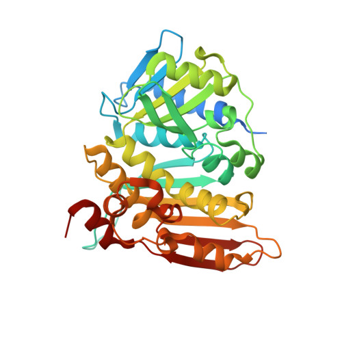
The crystal structure of methenyltetrahydromethanopterin cyclohydrolase from Methanobrevibacter ruminantium.
Carbone, V., Schofield, L.R., Beattie, A.K., Sutherland-Smith, A.J., Ronimus, R.S.(2013) Proteins 81: 2064-2070
- PubMed: 23873651
- DOI: https://doi.org/10.1002/prot.24372
- Primary Citation of Related Structures:
4FIO - PubMed Abstract:
Methenyltetrahydromethanopterin cyclohydrolase (Mch) is involved in the methanogenesis pathway of archaea as a C1 unit carrier where N(5) -formyl-tetrahydromethanopterin is converted to methenyl-tetrahydromethanopterin. Mch from Methanobrevibacter ruminantium was cloned, purified, crystallized and its crystal structure solved at 1.37 Å resolution. A biologically active trimer, the enzyme is composed of two domains including an N-terminal domain of six α-helices encompassing a series of four β-sheets and a predominantly anti-parallel β-sheet at the C-terminus flanked on one side by α-helices. Sequence and structural alignments have helped identify residues involved in substrate binding and trimer formation.
Organizational Affiliation:
AgResearch Limited, Grasslands Research Centre, Tennent Drive, Private Bag 11008, Palmerston North, 4442, New Zealand.


