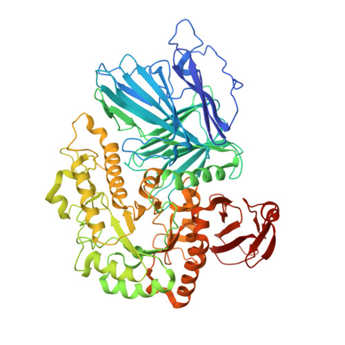
The molecular mechanism of the thermostable alpha-galactosidases AgaA and AgaB explained by X-ray crystallography and mutational studies
Merceron, R., Foucault, M., Haser, R., Mattes, R., Watzlawick, H., Gouet, P.(2012) J Biol Chem 287: 39642-39652
- PubMed: 23012371
- DOI: https://doi.org/10.1074/jbc.M112.394114
- Primary Citation of Related Structures:
4FNP, 4FNQ, 4FNR, 4FNS, 4FNT, 4FNU - PubMed Abstract:
The α-galactosidase AgaA from the thermophilic microorganism Geobacillus stearothermophilus has great industrial potential because it is fully active at 338 K against raffinose and can increase the yield of manufactured sucrose. AgaB has lower affinity for its natural substrates but is a powerful tool for the enzymatic synthesis of disaccharides by transglycosylation. These two enzymes have 97% identity and belong to the glycoside hydrolase (GH) family GH36 for which few structures are available. To understand the structural basis underlying the differences between these two enzymes, we determined the crystal structures of AgaA and AgaB by molecular replacement at 3.2- and 1.8 Å-resolution, respectively. We also solved a 2.8-Å structure of the AgaA(A355E) mutant, which has enzymatic properties similar to those of AgaB. We observe that residue 355 is located 20 Å away from the active site and that the A355E substitution causes structural rearrangements resulting in a significant displacement of the invariant Trp(336) at catalytic subsite -1. Hence, the active cleft of AgaA is narrowed in comparison with AgaB, and AgaA is more efficient than AgaB against its natural substrates. The structure of AgaA(A355E) complexed with 1-deoxygalactonojirimycin reveals an induced fit movement; there is a rupture of the electrostatic interaction between Glu(355) and Asn(335) and a return of Trp(336) to an optimal position for ligand stacking. The structures of two catalytic mutants of AgaA(A355E) complexed with raffinose and stachyose show that the binding interactions are stronger at subsite -1 to enable the binding of various α-galactosides.
Organizational Affiliation:
Biocrystallography and Structural Biology of Therapeutic Targets, Bases Moléculaires et Structurales des Systèmes Infectieux-Institut de Biologie et Chimie des Protéines, UMR 5086 CNRS, Université de Lyon 1, 7 passage du Vercors, 69367 Lyon Cedex 07, France.


