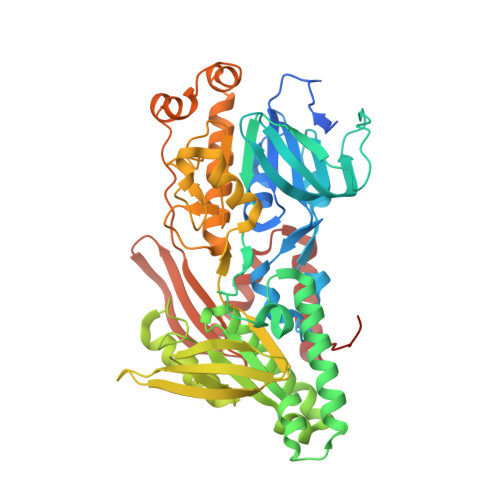
4G73

Crystal structure of NDH with NADH and Quinone


Experimental Data Snapshot

  • Method: X-RAY DIFFRACTION
  • Resolution: 2.52 Å
  • R-Value Free: 0.217 
  • R-Value Work: 0.189 
  • R-Value Observed: 0.190 

Starting Model: experimental
View more details

wwPDB Validation   3D Report Full Report


Ligand Structure Quality Assessment 


This is version 1.3 of the entry. See complete history


Literature

Structural insight into the type-II mitochondrial NADH dehydrogenases.

Feng, Y.Li, W.Li, J.Wang, J.Ge, J.Xu, D.Liu, Y.Wu, K.Zeng, Q.Wu, J.W.Tian, C.Zhou, B.Yang, M.

(2012) Nature 491: 478-482

  • DOI: https://doi.org/10.1038/nature11541
  • Primary Citation of Related Structures:  
    4G6G, 4G6H, 4G73, 4G74

  • PubMed Abstract: 

    The single-component type-II NADH dehydrogenases (NDH-2s) serve as alternatives to the multisubunit respiratory complex I (type-I NADH dehydrogenase (NDH-1), also called NADH:ubiquinone oxidoreductase; EC 1.6.5.3) in catalysing electron transfer from NADH to ubiquinone in the mitochondrial respiratory chain. The yeast NDH-2 (Ndi1) oxidizes NADH on the matrix side and reduces ubiquinone to maintain mitochondrial NADH/NAD(+) homeostasis. Ndi1 is a potential therapeutic agent for human diseases caused by complex I defects, particularly Parkinson's disease, because its expression restores the mitochondrial activity in animals with complex I deficiency. NDH-2s in pathogenic microorganisms are viable targets for new antibiotics. Here we solve the crystal structures of Ndi1 in its substrate-free, NADH-, ubiquinone- and NADH-ubiquinone-bound states, to help understand the catalytic mechanism of NDH-2s. We find that Ndi1 homodimerization through its carboxy-terminal domain is critical for its catalytic activity and membrane targeting. The structures reveal two ubiquinone-binding sites (UQ(I) and UQ(II)) in Ndi1. NADH and UQ(I) can bind to Ndi1 simultaneously to form a substrate-protein complex. We propose that UQ(I) interacts with FAD to act as an intermediate for electron transfer, and that NADH transfers electrons through this FAD-UQ(I) complex to UQ(II). Together our data reveal the regulatory and catalytic mechanisms of Ndi1 and may facilitate the development or targeting of NDH-2s for potential therapeutic applications.


  • Organizational Affiliation

    State Key Laboratory of Biomembrane and Membrane Biotechnology, Tsinghua-Peking Center for Life Sciences, School of Life Sciences, Tsinghua University, Beijing 100084, China.


Macromolecules
Find similar proteins by:  (by identity cutoff)  |  3D Structure
Entity ID: 1
MoleculeChains Sequence LengthOrganismDetailsImage
Rotenone-insensitive NADH-ubiquinone oxidoreductase, mitochondrial
A, B
502Saccharomyces cerevisiae S288CMutation(s): 0 
Gene Names: NDI1YML120CYM7056.06C
EC: 1.6.5.9
Membrane Entity: Yes 
UniProt
Find proteins for P32340 (Saccharomyces cerevisiae (strain ATCC 204508 / S288c))
Explore P32340 
Go to UniProtKB:  P32340
Entity Groups  
Sequence Clusters30% Identity50% Identity70% Identity90% Identity95% Identity100% Identity
UniProt GroupP32340
Sequence Annotations
Expand
  • Reference Sequence
Small Molecules
Ligands 4 Unique
IDChains Name / Formula / InChI Key2D Diagram3D Interactions
FAD
Query on FAD

Download Ideal Coordinates CCD File 
G [auth A],
R [auth B]
FLAVIN-ADENINE DINUCLEOTIDE
C27 H33 N9 O15 P2
VWWQXMAJTJZDQX-UYBVJOGSSA-N
NAI
Query on NAI

Download Ideal Coordinates CCD File 
L [auth A],
O [auth B]
1,4-DIHYDRONICOTINAMIDE ADENINE DINUCLEOTIDE
C21 H29 N7 O14 P2
BOPGDPNILDQYTO-NNYOXOHSSA-N
UQ5
Query on UQ5

Download Ideal Coordinates CCD File 
C [auth A],
D [auth A],
F [auth A],
P [auth B],
Q [auth B]
2,3-DIMETHOXY-5-METHYL-6-(3,11,15,19-TETRAMETHYL-EICOSA-2,6,10,14,18-PENTAENYL)-[1,4]BENZOQUINONE
C34 H50 O4
NYFAQDMDAFCWPU-UVCHAVPFSA-N
MG
Query on MG

Download Ideal Coordinates CCD File 
E [auth A]
H [auth A]
I [auth A]
J [auth A]
K [auth A]
E [auth A],
H [auth A],
I [auth A],
J [auth A],
K [auth A],
M [auth B],
N [auth B],
S [auth B],
T [auth B],
U [auth B],
V [auth B]
MAGNESIUM ION
Mg
JLVVSXFLKOJNIY-UHFFFAOYSA-N
Experimental Data & Validation

Experimental Data

  • Method: X-RAY DIFFRACTION
  • Resolution: 2.52 Å
  • R-Value Free: 0.217 
  • R-Value Work: 0.189 
  • R-Value Observed: 0.190 
  • Space Group: C 2 2 21
Unit Cell:
Length ( Å )Angle ( ˚ )
a = 127.406α = 90
b = 230.186β = 90
c = 112.921γ = 90
Software Package:
Software NamePurpose
HKL-2000data collection
PHASESphasing
PHENIXrefinement
HKL-2000data reduction
HKL-2000data scaling

Structure Validation

View Full Validation Report


Ligand Structure Quality Assessment 


Entry History 

Deposition Data

Revision History  (Full details and data files)

  • Version 1.0: 2012-10-24
    Type: Initial release
  • Version 1.1: 2012-12-05
    Changes: Database references
  • Version 1.2: 2013-11-06
    Changes: Non-polymer description
  • Version 1.3: 2023-11-08
    Changes: Data collection, Database references, Derived calculations, Refinement description