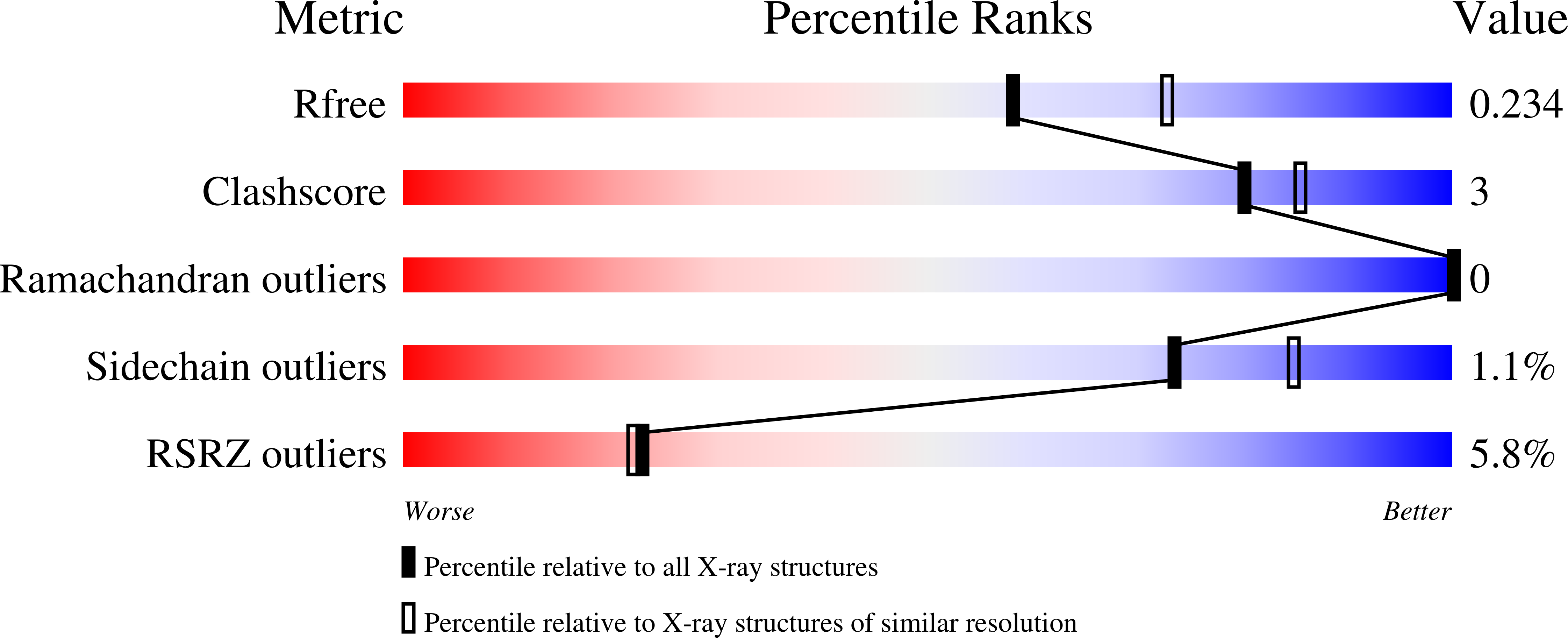
4GKA

Crystal structure of purine nucleoside phosphorylase (W16Y, W94Y, W178Y, H257W) mutant from human complexed with phosphate


Experimental Data Snapshot

  • Method: X-RAY DIFFRACTION
  • Resolution: 2.20 Å
  • R-Value Free: 0.232 
  • R-Value Work: 0.188 
  • R-Value Observed: 0.190 

wwPDB Validation   3D Report Full Report


This is version 1.4 of the entry. See complete history


Literature

Catalytic Site Conformations in Human PNP by (19)F-NMR and Crystallography.

Suarez, J.Haapalainen, A.M.Cahill, S.M.Ho, M.C.Yan, F.Almo, S.C.Schramm, V.L.

(2013) Chem Biol 20: 212-222

  • DOI: https://doi.org/10.1016/j.chembiol.2013.01.009
  • Primary Citation of Related Structures:  
    4EAR, 4EB8, 4ECE, 4GKA

  • PubMed Abstract: 

    Purine nucleoside phosphorylase (PNP) is a target for leukemia, gout, and autoimmune disorders. Dynamic motion of catalytic site loops has been implicated in catalysis, but experimental evidence was lacking. We replaced catalytic site groups His257 or His64 with 6-fluoro-tryptophan (6FW) as site-specific NMR probes. Conformational adjustments in the 6FW-His257-helical and His64-6FW-loop regions were characterized in PNP phosphate-bound enzyme and in complexes with catalytic site ligands, including transition state analogs. Chemical shift and line-shape changes associated with these complexes revealed dynamic coexistence of several conformational states in these regions in phosphate-bound enzyme and altered or single conformations in other complexes. These conformations were also characterized by X-ray crystallography. Specific (19)F-Trp labels and X-ray crystallography provide multidimensional characterization of conformational states for free, catalytic, and inhibited complexes of human PNP.


  • Organizational Affiliation

    Department of Biochemistry, Albert Einstein College of Medicine of Yeshiva University, 1300 Morris Park Avenue, Bronx, NY 10461, USA.


Macromolecules
Find similar proteins by:  (by identity cutoff)  |  3D Structure
Entity ID: 1
MoleculeChains Sequence LengthOrganismDetailsImage
Purine nucleoside phosphorylase
A, B, C, D, E
A, B, C, D, E, F
324Homo sapiensMutation(s): 5 
Gene Names: NPPNPX00737.1
EC: 2.4.2.1
UniProt & NIH Common Fund Data Resources
Find proteins for P00491 (Homo sapiens)
Explore P00491 
Go to UniProtKB:  P00491
PHAROS:  P00491
GTEx:  ENSG00000198805 
Entity Groups  
Sequence Clusters30% Identity50% Identity70% Identity90% Identity95% Identity100% Identity
UniProt GroupP00491
Sequence Annotations
Expand
  • Reference Sequence
Small Molecules
Ligands 2 Unique
IDChains Name / Formula / InChI Key2D Diagram3D Interactions
PO4
Query on PO4

Download Ideal Coordinates CCD File 
G [auth A]
H [auth B]
I [auth C]
K [auth D]
L [auth E]
G [auth A],
H [auth B],
I [auth C],
K [auth D],
L [auth E],
M [auth F]
PHOSPHATE ION
O4 P
NBIIXXVUZAFLBC-UHFFFAOYSA-K
GOL
Query on GOL

Download Ideal Coordinates CCD File 
J [auth C],
N [auth F]
GLYCEROL
C3 H8 O3
PEDCQBHIVMGVHV-UHFFFAOYSA-N
Experimental Data & Validation

Experimental Data

  • Method: X-RAY DIFFRACTION
  • Resolution: 2.20 Å
  • R-Value Free: 0.232 
  • R-Value Work: 0.188 
  • R-Value Observed: 0.190 
  • Space Group: P 1 21 1
Unit Cell:
Length ( Å )Angle ( ˚ )
a = 53.882α = 90
b = 233.67β = 97.33
c = 70.077γ = 90
Software Package:
Software NamePurpose
ADSCdata collection
MOLREPphasing
REFMACrefinement
HKL-3000data reduction
SCALEPACKdata scaling

Structure Validation

View Full Validation Report


Entry History 

Deposition Data

Revision History  (Full details and data files)

  • Version 1.0: 2013-02-06
    Type: Initial release
  • Version 1.1: 2013-03-06
    Changes: Database references
  • Version 1.2: 2013-03-13
    Changes: Database references
  • Version 1.3: 2022-12-21
    Changes: Database references, Derived calculations
  • Version 1.4: 2024-05-22
    Changes: Data collection