4GYJ

Crystal structure of mutant (D318N) bacillus subtilis family 3 glycoside hydrolase (nagz) in complex with glcnac-murnac (space group P1)


Experimental Data Snapshot

  • Method: X-RAY DIFFRACTION
  • Resolution: 1.65 Å
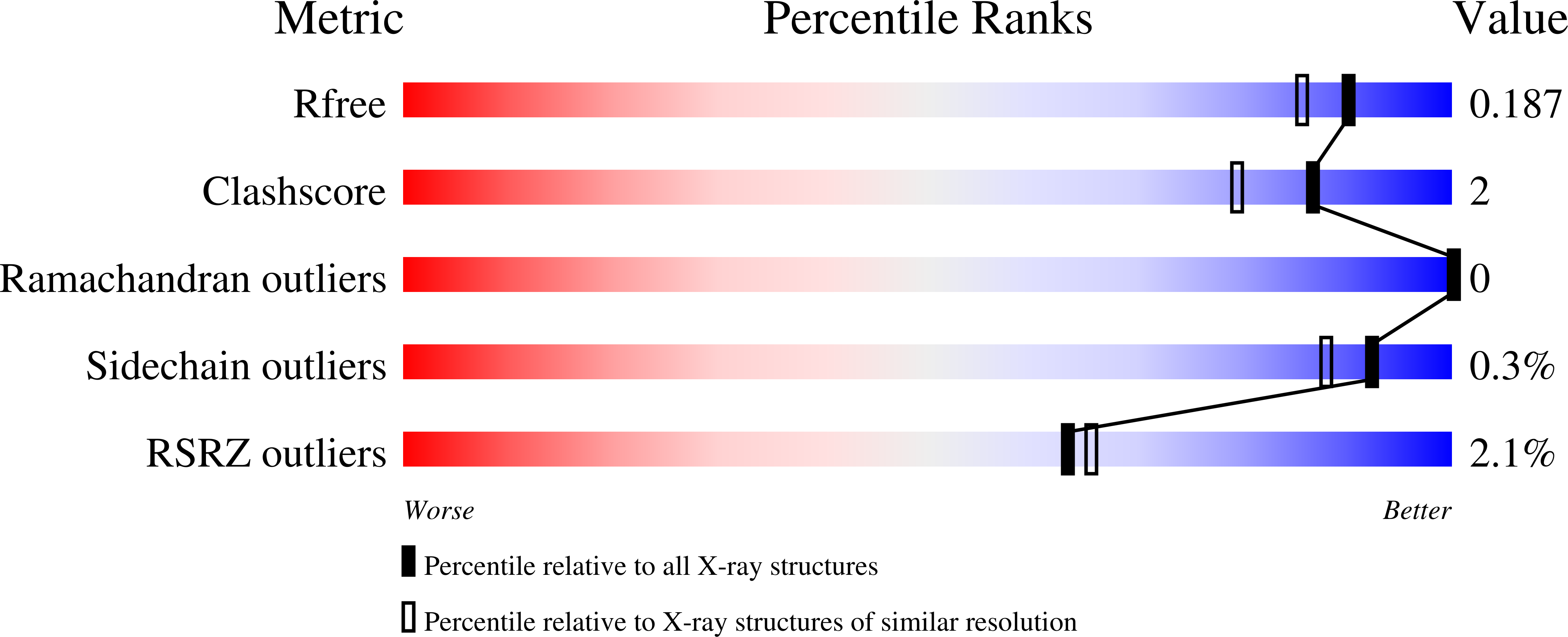
  • R-Value Free: 0.190 
  • R-Value Work: 0.163 
  • R-Value Observed: 0.164 

Starting Model: experimental
View more details

wwPDB Validation   3D Report Full Report


Ligand Structure Quality Assessment 


This is version 2.1 of the entry. See complete history


Literature

Active Site Plasticity within the Glycoside Hydrolase NagZ Underlies a Dynamic Mechanism of Substrate Distortion.

Bacik, J.P.Whitworth, G.E.Stubbs, K.A.Vocadlo, D.J.Mark, B.L.

(2012) Chem Biol 19: 1471-1482

  • DOI: https://doi.org/10.1016/j.chembiol.2012.09.016
  • Primary Citation of Related Structures:  
    4GVF, 4GVG, 4GVH, 4GVI, 4GYJ, 4GYK

  • PubMed Abstract: 

    NagZ is a glycoside hydrolase that participates in peptidoglycan (PG) recycling by removing β-N-acetylglucosamine from PG fragments that are excised from the bacterial cell wall during growth. Notably, the products formed by NagZ, 1,6-anhydroMurNAc-peptides, activate β-lactam resistance in many Gram-negative bacteria, making this enzyme of interest as a potential therapeutic target. Crystal structure determinations of NagZ from Salmonella typhimurium and Bacillus subtilis in complex with natural substrate, trapped as a glycosyl-enzyme intermediate, and bound to product, define the reaction coordinate of the NagZ family of enzymes. The structures, combined with kinetic studies, reveal an uncommon degree of structural plasticity within the active site of a glycoside hydrolase, and unveil how NagZ drives substrate distortion using a highly mobile loop that contains a conserved histidine that has been proposed as the general acid/base.


  • Organizational Affiliation

    Department of Microbiology, University of Manitoba, Winnipeg, MB R3T 2N2, Canada.


Macromolecules
Find similar proteins by:  (by identity cutoff)  |  3D Structure
Entity ID: 1
MoleculeChains Sequence LengthOrganismDetailsImage
Uncharacterized lipoprotein ybbD
A, B
648Bacillus subtilis subsp. subtilis str. 168Mutation(s): 1 
Gene Names: BSU01660NagZybbDyzbA
EC: 3.2.1.52
UniProt
Find proteins for P40406 (Bacillus subtilis (strain 168))
Explore P40406 
Go to UniProtKB:  P40406
Entity Groups  
Sequence Clusters30% Identity50% Identity70% Identity90% Identity95% Identity100% Identity
UniProt GroupP40406
Sequence Annotations
Expand
  • Reference Sequence
Oligosaccharides

Help

Entity ID: 2
MoleculeChains Length2D Diagram Glycosylation3D Interactions
2-acetamido-2-deoxy-beta-D-glucopyranose-(1-4)-N-acetyl-alpha-muramic acid
C, D
2N/A
Glycosylation Resources
GlyTouCan:  G88499BX
GlyCosmos:  G88499BX
Experimental Data & Validation

Experimental Data

  • Method: X-RAY DIFFRACTION
  • Resolution: 1.65 Å
  • R-Value Free: 0.190 
  • R-Value Work: 0.163 
  • R-Value Observed: 0.164 
  • Space Group: P 1
Unit Cell:
Length ( Å )Angle ( ˚ )
a = 58.45α = 98.65
b = 73.39β = 110.14
c = 83.43γ = 92.43
Software Package:
Software NamePurpose
PHENIXrefinement
MxDCdata collection
PHASERphasing
MOSFLMdata reduction
SCALAdata scaling

Structure Validation

View Full Validation Report


Ligand Structure Quality Assessment 


Entry History 

Deposition Data

Revision History  (Full details and data files)

  • Version 1.0: 2012-12-19
    Type: Initial release
  • Version 1.1: 2014-10-08
    Changes: Structure summary
  • Version 1.2: 2015-01-28
    Changes: Derived calculations
  • Version 2.0: 2020-07-29
    Type: Remediation
    Reason: Carbohydrate remediation
    Changes: Advisory, Atomic model, Data collection, Database references, Derived calculations, Refinement description, Structure summary
  • Version 2.1: 2023-09-13
    Changes: Data collection, Database references, Refinement description, Structure summary