4H53

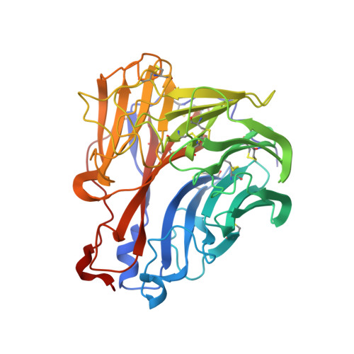
Influenza N2-Tyr406Asp neuraminidase in complex with beta-Neu5Ac


Experimental Data Snapshot

  • Method: X-RAY DIFFRACTION
  • Resolution: 1.50 Å
  • R-Value Free: 0.161 
  • R-Value Work: 0.140 
  • R-Value Observed: 0.141 

Starting Model: experimental
View more details

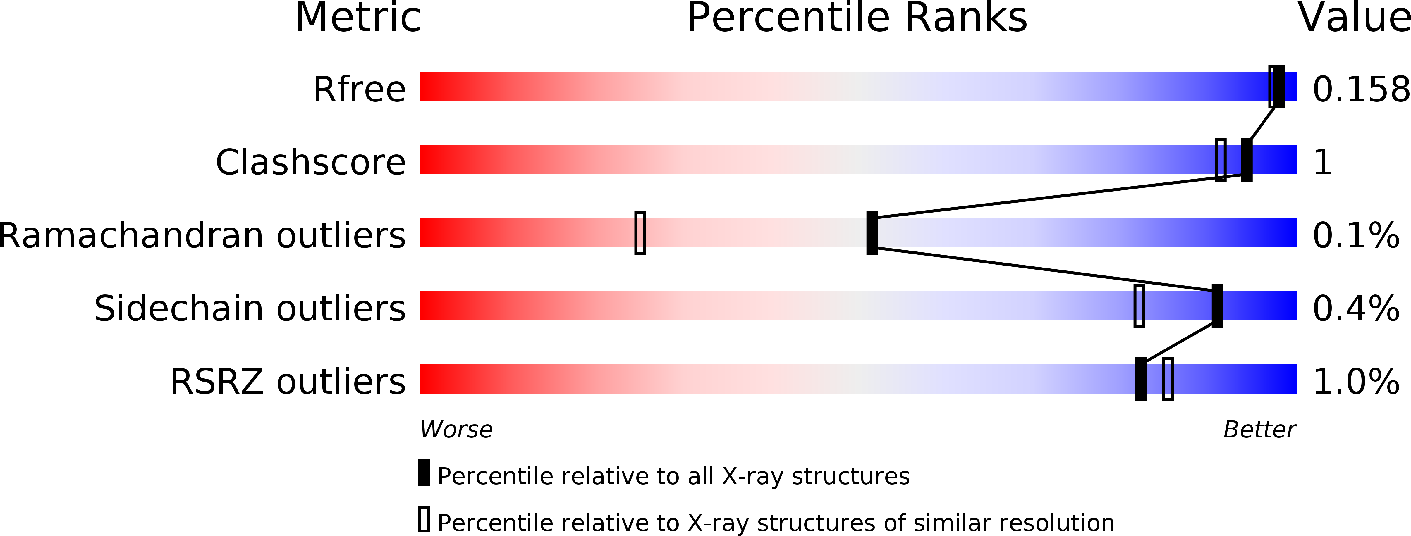
wwPDB Validation   3D Report Full Report


Ligand Structure Quality Assessment 


This is version 2.1 of the entry. See complete history


Literature

Influenza neuraminidase operates via a nucleophilic mechanism and can be targeted by covalent inhibitors

Vavricka, C.J.Liu, Y.Kiyota, H.Sriwilaijaroen, N.Qi, J.Tanaka, K.Wu, Y.Li, Q.Li, Y.Yan, J.Suzuki, Y.Gao, G.F.

(2013) Nat Commun 4: 1491-1491

  • DOI: https://doi.org/10.1038/ncomms2487
  • Primary Citation of Related Structures:  
    4H52, 4H53

  • PubMed Abstract: 

    Development of novel influenza neuraminidase inhibitors is critical for preparedness against influenza outbreaks. Knowledge of the neuraminidase enzymatic mechanism and transition-state analogue, 2-deoxy-2,3-didehydro-N-acetylneuraminic acid, contributed to the development of the first generation anti-neuraminidase drugs, zanamivir and oseltamivir. However, lack of evidence regarding influenza neuraminidase key catalytic residues has limited strategies for novel neuraminidase inhibitor design. Here, we confirm that influenza neuraminidase conserved Tyr406 is the key catalytic residue that may function as a nucleophile; thus, mechanism-based covalent inhibition of influenza neuraminidase was conceived. Crystallographic studies reveal that 2α,3ax-difluoro-N-acetylneuraminic acid forms a covalent bond with influenza neuraminidase Tyr406 and the compound was found to possess potent anti-influenza activity against both influenza A and B viruses. Our results address many unanswered questions about the influenza neuraminidase catalytic mechanism and demonstrate that covalent inhibition of influenza neuraminidase is a promising and novel strategy for the development of next-generation influenza drugs.


  • Organizational Affiliation

    Research Network of Immunity and Health, Beijing Institutes of Life Science, Beijing 100101, China.


Macromolecules
Find similar proteins by:  (by identity cutoff)  |  3D Structure
Entity ID: 1
MoleculeChains Sequence LengthOrganismDetailsImage
Neuraminidase
A, B, C, D
388Influenza A virus (A/RI/5+/1957(H2N2))Mutation(s): 1 
EC: 3.2.1.18
UniProt
Find proteins for Q194T1 (Influenza A virus)
Explore Q194T1 
Go to UniProtKB:  Q194T1
Entity Groups  
Sequence Clusters30% Identity50% Identity70% Identity90% Identity95% Identity100% Identity
UniProt GroupQ194T1
Glycosylation
Glycosylation Sites: 2
Sequence Annotations
Expand
  • Reference Sequence
Oligosaccharides

Help

Entity ID: 2
MoleculeChains Length2D Diagram Glycosylation3D Interactions
alpha-D-mannopyranose-(1-3)-[alpha-D-mannopyranose-(1-6)]alpha-D-mannopyranose-(1-6)-[alpha-D-mannopyranose-(1-3)]beta-D-mannopyranose-(1-4)-2-acetamido-2-deoxy-beta-D-glucopyranose-(1-4)-2-acetamido-2-deoxy-beta-D-glucopyranose
E
7N-Glycosylation
Glycosylation Resources
GlyTouCan:  G55220VL
GlyCosmos:  G55220VL
GlyGen:  G55220VL
Entity ID: 3
MoleculeChains Length2D Diagram Glycosylation3D Interactions
N-acetyl-alpha-neuraminic acid-(2-3)-beta-D-galactopyranose
F, I, L
2N/A
Glycosylation Resources
GlyTouCan:  G30207PZ
GlyCosmos:  G30207PZ
GlyGen:  G30207PZ
Entity ID: 4
MoleculeChains Length2D Diagram Glycosylation3D Interactions
alpha-D-mannopyranose-(1-3)-beta-D-mannopyranose-(1-4)-2-acetamido-2-deoxy-beta-D-glucopyranose-(1-4)-2-acetamido-2-deoxy-beta-D-glucopyranose
G, H, J, K
4N-Glycosylation
Glycosylation Resources
GlyTouCan:  G81315DD
GlyCosmos:  G81315DD
GlyGen:  G81315DD
Entity ID: 5
MoleculeChains Length2D Diagram Glycosylation3D Interactions
2-acetamido-2-deoxy-beta-D-glucopyranose-(1-4)-2-acetamido-2-deoxy-beta-D-glucopyranose
M
2N-Glycosylation
Glycosylation Resources
GlyTouCan:  G42666HT
GlyCosmos:  G42666HT
GlyGen:  G42666HT
Entity ID: 6
MoleculeChains Length2D Diagram Glycosylation3D Interactions
alpha-D-mannopyranose-(1-3)-alpha-D-mannopyranose-(1-6)-[alpha-D-mannopyranose-(1-3)]beta-D-mannopyranose-(1-4)-2-acetamido-2-deoxy-beta-D-glucopyranose-(1-4)-2-acetamido-2-deoxy-beta-D-glucopyranose
N
6N-Glycosylation
Glycosylation Resources
GlyTouCan:  G09724ZC
GlyCosmos:  G09724ZC
GlyGen:  G09724ZC
Small Molecules
Ligands 4 Unique
IDChains Name / Formula / InChI Key2D Diagram3D Interactions
SLB
Query on SLB

Download Ideal Coordinates CCD File 
Q [auth A],
S [auth B],
U [auth C]
N-acetyl-beta-neuraminic acid
C11 H19 N O9
SQVRNKJHWKZAKO-PFQGKNLYSA-N
NAG
Query on NAG

Download Ideal Coordinates CCD File 
P [auth A]2-acetamido-2-deoxy-beta-D-glucopyranose
C8 H15 N O6
OVRNDRQMDRJTHS-FMDGEEDCSA-N
GOL
Query on GOL

Download Ideal Coordinates CCD File 
V [auth D]GLYCEROL
C3 H8 O3
PEDCQBHIVMGVHV-UHFFFAOYSA-N
CA
Query on CA

Download Ideal Coordinates CCD File 
O [auth A],
R [auth B],
T [auth C]
CALCIUM ION
Ca
BHPQYMZQTOCNFJ-UHFFFAOYSA-N
Experimental Data & Validation

Experimental Data

  • Method: X-RAY DIFFRACTION
  • Resolution: 1.50 Å
  • R-Value Free: 0.161 
  • R-Value Work: 0.140 
  • R-Value Observed: 0.141 
  • Space Group: P 1 21 1
Unit Cell:
Length ( Å )Angle ( ˚ )
a = 83.217α = 90
b = 114.831β = 99.71
c = 84.259γ = 90
Software Package:
Software NamePurpose
HKL-2000data collection
PHASESphasing
PHENIXrefinement
HKL-2000data reduction
HKL-2000data scaling

Structure Validation

View Full Validation Report


Ligand Structure Quality Assessment 


Entry History 

Deposition Data

Revision History  (Full details and data files)

  • Version 1.0: 2013-02-20
    Type: Initial release
  • Version 1.1: 2013-02-27
    Changes: Structure summary
  • Version 1.2: 2013-07-31
    Changes: Database references, Derived calculations
  • Version 2.0: 2020-07-29
    Type: Remediation
    Reason: Carbohydrate remediation
    Changes: Advisory, Atomic model, Data collection, Database references, Derived calculations, Structure summary
  • Version 2.1: 2023-11-08
    Changes: Data collection, Database references, Refinement description, Structure summary