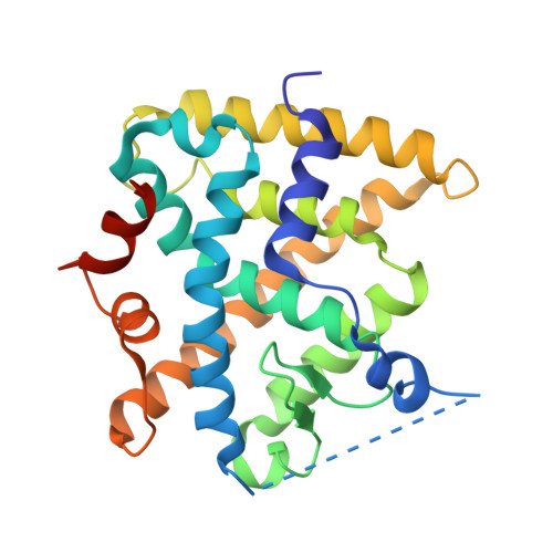
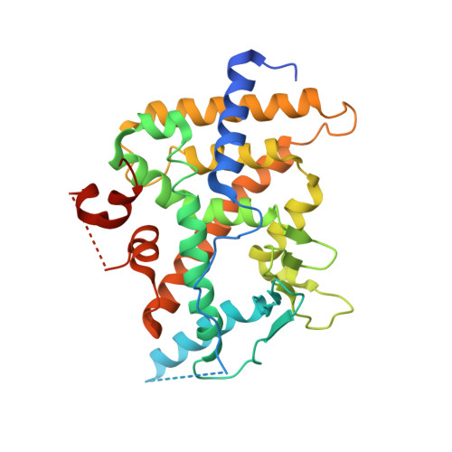
4J5X

Crystal Structure of the SR12813-bound PXR/RXRalpha LBD Heterotetramer Complex


Experimental Data Snapshot

  • Method: X-RAY DIFFRACTION
  • Resolution: 2.80 Å
  • R-Value Free: 0.298 
  • R-Value Work: 0.245 
  • R-Value Observed: 0.248 

wwPDB Validation   3D Report Full Report


Ligand Structure Quality Assessment 


This is version 1.3 of the entry. See complete history


Literature

Structural and Functional Analysis of the Human Nuclear Xenobiotic Receptor PXR in Complex with RXRalpha.

Wallace, B.D.Betts, L.Talmage, G.Pollet, R.M.Holman, N.S.Redinbo, M.R.

(2013) J Mol Biol 425: 2561-2577

  • DOI: https://doi.org/10.1016/j.jmb.2013.04.012
  • Primary Citation of Related Structures:  
    4J5W, 4J5X

  • PubMed Abstract: 

    The human nuclear xenobiotic receptor PXR recognizes a range of potentially harmful drugs and endobiotic chemicals but must complex with the nuclear receptor RXRα to control the expression of numerous drug metabolism genes. To date, the structural basis and functional consequences of this interaction have remained unclear. Here we present 2.8-Å-resolution crystal structures of the heterodimeric complex formed between the ligand-binding domains of human PXR and RXRα. These structures establish that PXR and RXRα form a heterotetramer unprecedented in the nuclear receptor family of ligand-regulated transcription factors. We further show that both PXR and RXRα bind to the transcriptional coregulator SRC-1 with higher affinity when they are part of the PXR/RXRα heterotetramer complex than they do when each ligand-binding domain is examined alone. Furthermore, we purify the full-length forms of each receptor from recombinant bacterial expression systems and characterize their interactions with a range of direct and everted repeat DNA elements. Taken together, these data advance our understanding of PXR, the master regulator of drug metabolism gene expression in humans, in its functional partnership with RXRα.


  • Organizational Affiliation

    Department of Chemistry, Biochemistry and Microbiology, University of North Carolina at Chapel Hill, 250 Bell Tower Drive, Chapel Hill, NC 27599, USA.


Macromolecules
Find similar proteins by:  (by identity cutoff)  |  3D Structure
Entity ID: 1
MoleculeChains Sequence LengthOrganismDetailsImage
Retinoic acid receptor RXR-alpha, Nuclear receptor coactivator 1A [auth D],
B [auth C]
264Homo sapiensMutation(s): 0 
Gene Names: NR2B1RXRABHLHE74NCOA1SRC1
EC: 2.3.1.48
UniProt & NIH Common Fund Data Resources
Find proteins for Q15788 (Homo sapiens)
Explore Q15788 
Go to UniProtKB:  Q15788
PHAROS:  Q15788
GTEx:  ENSG00000084676 
Find proteins for P19793 (Homo sapiens)
Explore P19793 
Go to UniProtKB:  P19793
PHAROS:  P19793
GTEx:  ENSG00000186350 
Entity Groups  
Sequence Clusters30% Identity50% Identity70% Identity90% Identity95% Identity100% Identity
UniProt GroupsQ15788P19793
Sequence Annotations
Expand
  • Reference Sequence
Find similar proteins by:  (by identity cutoff)  |  3D Structure
Entity ID: 2
MoleculeChains Sequence LengthOrganismDetailsImage
Nuclear receptor subfamily 1 group I member 2, Nuclear receptor coactivator 1C [auth A],
D [auth B]
336Homo sapiensMutation(s): 0 
Gene Names: NR1I2PXRBHLHE74NCOA1SRC1
EC: 2.3.1.48
UniProt & NIH Common Fund Data Resources
Find proteins for Q15788 (Homo sapiens)
Explore Q15788 
Go to UniProtKB:  Q15788
PHAROS:  Q15788
GTEx:  ENSG00000084676 
Find proteins for O75469 (Homo sapiens)
Explore O75469 
Go to UniProtKB:  O75469
PHAROS:  O75469
GTEx:  ENSG00000144852 
Entity Groups  
Sequence Clusters30% Identity50% Identity70% Identity90% Identity95% Identity100% Identity
UniProt GroupsQ15788O75469
Sequence Annotations
Expand
  • Reference Sequence
Small Molecules
Ligands 1 Unique
IDChains Name / Formula / InChI Key2D Diagram3D Interactions
SRL
Query on SRL

Download Ideal Coordinates CCD File 
E [auth A],
F [auth B]
[2-(3,5-DI-TERT-BUTYL-4-HYDROXY-PHENYL)-1-(DIETHOXY-PHOSPHORYL)-VINYL]-PHOSPHONIC ACID DIETHLYL ESTER
C24 H42 O7 P2
YQLJDECYQDRSBI-UHFFFAOYSA-N
Experimental Data & Validation

Experimental Data

  • Method: X-RAY DIFFRACTION
  • Resolution: 2.80 Å
  • R-Value Free: 0.298 
  • R-Value Work: 0.245 
  • R-Value Observed: 0.248 
  • Space Group: P 21 21 21
Unit Cell:
Length ( Å )Angle ( ˚ )
a = 70.09α = 90
b = 120.3β = 90
c = 175.79γ = 90
Software Package:
Software NamePurpose
PHENIXrefinement
PDB_EXTRACTdata extraction
JBluIce-EPICSdata collection
XDSdata reduction
XSCALEdata scaling
PHASERphasing

Structure Validation

View Full Validation Report


Ligand Structure Quality Assessment 


Entry History 

Deposition Data

Revision History  (Full details and data files)

  • Version 1.0: 2013-08-21
    Type: Initial release
  • Version 1.1: 2017-08-16
    Changes: Source and taxonomy
  • Version 1.2: 2017-11-15
    Changes: Refinement description
  • Version 1.3: 2024-02-28
    Changes: Data collection, Database references, Derived calculations