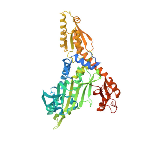
4K88

Crystal structure of human prolyl-tRNA synthetase (halofuginone bound form)


Experimental Data Snapshot

  • Method: X-RAY DIFFRACTION
  • Resolution: 2.62 Å
  • R-Value Free: 0.276 
  • R-Value Work: 0.237 
  • R-Value Observed: 0.239 

wwPDB Validation   3D Report Full Report


Ligand Structure Quality Assessment 


This is version 1.2 of the entry. See complete history


Literature

Conformational changes in human prolyl-tRNA synthetase upon binding of the substrates proline and ATP and the inhibitor halofuginone.

Son, J.Lee, E.H.Park, M.Kim, J.H.Kim, J.Kim, S.Jeon, Y.H.Hwang, K.Y.

(2013) Acta Crystallogr D Biol Crystallogr 69: 2136-2145

  • DOI: https://doi.org/10.1107/S0907444913020556
  • Primary Citation of Related Structures:  
    4K86, 4K87, 4K88

  • PubMed Abstract: 

    Aminoacyl-tRNA synthetases recognize cognate amino acids and tRNAs from their noncognate counterparts and catalyze the formation of aminoacyl-tRNAs. Halofuginone (HF), a coccidiostat used in veterinary medicine, exerts its effects by acting as a high-affinity inhibitor of the enzyme glutamyl-prolyl-tRNA synthetase (EPRS). In order to elucidate the precise molecular basis of this inhibition mechanism of human EPRS, the crystal structures of the prolyl-tRNA synthetase domain of human EPRS (hPRS) at 2.4 Å resolution (hPRS-apo), of hPRS complexed with ATP and the substrate proline at 2.3 Å resolution (hPRS-sub) and of hPRS complexed with HF at 2.62 Å resolution (hPRS-HF) are presented. These structures show plainly that motif 1 functions as a cap in hPRS, which is loosely opened in hPRS-apo, tightly closed in hPRS-sub and incorrectly closed in hPRS-HF. In addition, the structural analyses are consistent with more effective binding of hPRS to HF with ATP. Mutagenesis and biochemical analysis confirmed the key roles of two residues, Phe1097 and Arg1152, in the HF inhibition mechanism. These structures will lead to the development of more potent and selective hPRS inhibitors for promoting inflammatory resolution.


  • Organizational Affiliation

    Department of Biosystems and Biotechnology, Korea University, Anam-dong 5, Seongbuk-gu, Seoul 136-701, Republic of Korea.


Macromolecules
Find similar proteins by:  (by identity cutoff)  |  3D Structure
Entity ID: 1
MoleculeChains Sequence LengthOrganismDetailsImage
Proline--tRNA ligase535Homo sapiensMutation(s): 0 
Gene Names: EPRS
EC: 6.1.1.15 (PDB Primary Data), 6.1.1.17 (UniProt)
UniProt & NIH Common Fund Data Resources
Find proteins for P07814 (Homo sapiens)
Explore P07814 
Go to UniProtKB:  P07814
PHAROS:  P07814
GTEx:  ENSG00000136628 
Entity Groups  
Sequence Clusters30% Identity50% Identity70% Identity90% Identity95% Identity100% Identity
UniProt GroupP07814
Sequence Annotations
Expand
  • Reference Sequence
Small Molecules
Ligands 2 Unique
IDChains Name / Formula / InChI Key2D Diagram3D Interactions
HFG
Query on HFG

Download Ideal Coordinates CCD File 
C [auth A]7-bromo-6-chloro-3-{3-[(2R,3S)-3-hydroxypiperidin-2-yl]-2-oxopropyl}quinazolin-4(3H)-one
C16 H17 Br Cl N3 O3
LVASCWIMLIKXLA-CABCVRRESA-N
ZN
Query on ZN

Download Ideal Coordinates CCD File 
B [auth A]ZINC ION
Zn
PTFCDOFLOPIGGS-UHFFFAOYSA-N
Experimental Data & Validation

Experimental Data

  • Method: X-RAY DIFFRACTION
  • Resolution: 2.62 Å
  • R-Value Free: 0.276 
  • R-Value Work: 0.237 
  • R-Value Observed: 0.239 
  • Space Group: P 31 2 1
Unit Cell:
Length ( Å )Angle ( ˚ )
a = 120.813α = 90
b = 120.813β = 90
c = 104.711γ = 120
Software Package:
Software NamePurpose
PHENIXrefinement
PDB_EXTRACTdata extraction
HKL-2000data collection
HKL-2000data reduction
HKL-2000data scaling
PHENIXphasing

Structure Validation

View Full Validation Report


Ligand Structure Quality Assessment 


Entry History 

Deposition Data

Revision History  (Full details and data files)

  • Version 1.0: 2013-10-09
    Type: Initial release
  • Version 1.1: 2019-12-25
    Changes: Database references
  • Version 1.2: 2024-03-20
    Changes: Data collection, Database references, Derived calculations