4KXM

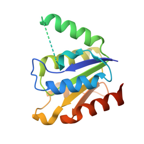
Crystal structure of DNPH1 (RCL) WITH N6-ISOPENTENYL-AMP


Experimental Data Snapshot

  • Method: X-RAY DIFFRACTION
  • Resolution: 2.24 Å
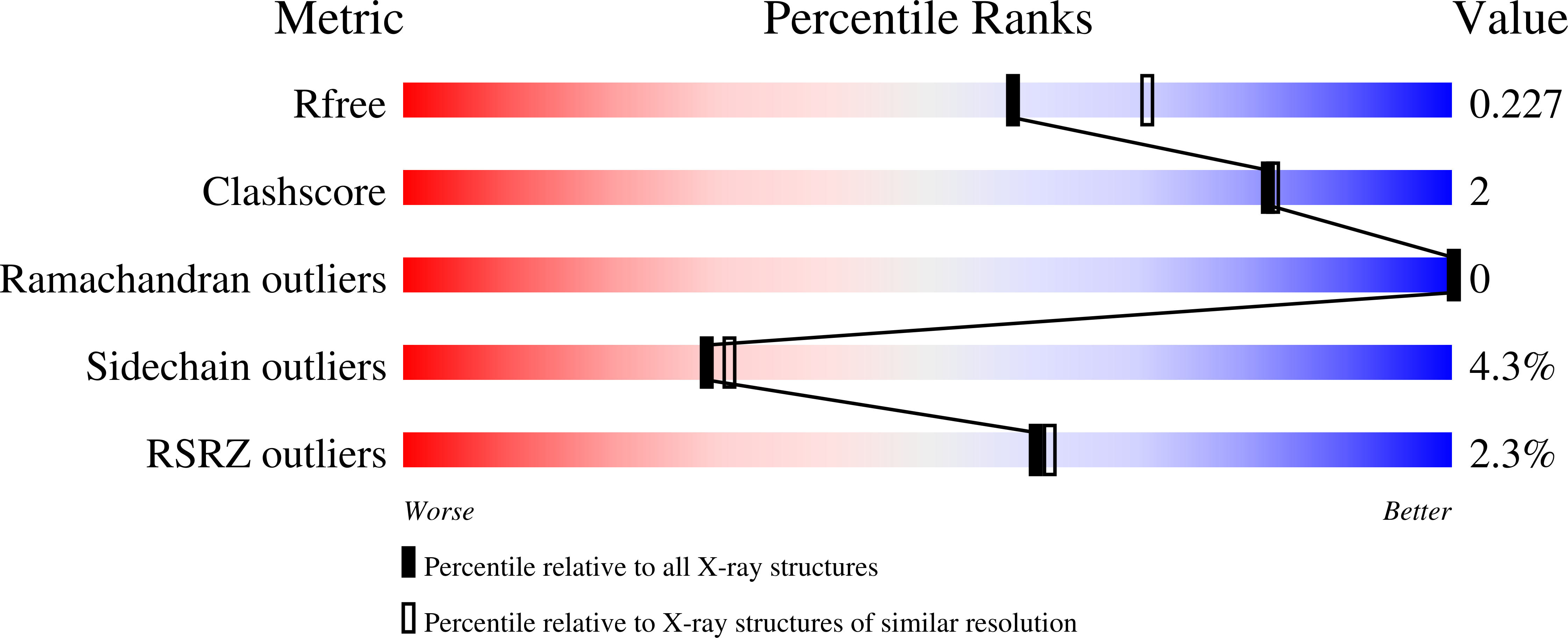
  • R-Value Free: 0.227 
  • R-Value Work: 0.174 
  • R-Value Observed: 0.176 

Starting Model: experimental
View more details

wwPDB Validation   3D Report Full Report


Ligand Structure Quality Assessment 


This is version 1.1 of the entry. See complete history


Literature

N (6)-substituted AMPs inhibit mammalian deoxynucleotide N-hydrolase DNPH1.

Amiable, C.Pochet, S.Padilla, A.Labesse, G.Kaminski, P.A.

(2013) PLoS One 8: e80755-e80755

  • DOI: https://doi.org/10.1371/journal.pone.0080755
  • Primary Citation of Related Structures:  
    4KXL, 4KXM, 4KXN

  • PubMed Abstract: 

    The gene dnph1 (or rcl) encodes a hydrolase that cleaves the 2'-deoxyribonucleoside 5'-monophosphate (dNMP) N-glycosidic bond to yield a free nucleobase and 2-deoxyribose 5-phosphate. Recently, the crystal structure of rat DNPH1, a potential target for anti-cancer therapies, suggested that various analogs of AMP may inhibit this enzyme. From this result, we asked whether N (6)-substituted AMPs, and among them, cytotoxic cytokinin riboside 5'-monophosphates, may inhibit DNPH1. Here, we characterized the structural and thermodynamic aspects of the interactions of these various analogs with DNPH1. Our results indicate that DNPH1 is inhibited by cytotoxic cytokinins at concentrations that inhibit cell growth.


  • Organizational Affiliation

    Institut Pasteur, Unité de Chimie et Biocatalyse, Paris, France ; CNRS, UMR3523, Paris, France ; Université Paris Descartes Sorbonne Paris Cité, Paris, France.


Macromolecules
Find similar proteins by:  (by identity cutoff)  |  3D Structure
Entity ID: 1
MoleculeChains Sequence LengthOrganismDetailsImage
2'-deoxynucleoside 5'-phosphate N-hydrolase 1
A, B, C, D
152Rattus norvegicusMutation(s): 1 
Gene Names: Dnph1Rcl
EC: 3.2.2
UniProt
Find proteins for O35820 (Rattus norvegicus)
Explore O35820 
Go to UniProtKB:  O35820
Entity Groups  
Sequence Clusters30% Identity50% Identity70% Identity90% Identity95% Identity100% Identity
UniProt GroupO35820
Sequence Annotations
Expand
  • Reference Sequence
Experimental Data & Validation

Experimental Data

  • Method: X-RAY DIFFRACTION
  • Resolution: 2.24 Å
  • R-Value Free: 0.227 
  • R-Value Work: 0.174 
  • R-Value Observed: 0.176 
  • Space Group: P 1 21 1
Unit Cell:
Length ( Å )Angle ( ˚ )
a = 32.146α = 90
b = 96.568β = 101.58
c = 79.034γ = 90
Software Package:
Software NamePurpose
SCALAdata scaling
MOLREPphasing
REFMACrefinement
PDB_EXTRACTdata extraction
MOSFLMdata reduction

Structure Validation

View Full Validation Report


Ligand Structure Quality Assessment 


Entry History 

Deposition Data

Revision History  (Full details and data files)

  • Version 1.0: 2014-02-05
    Type: Initial release
  • Version 1.1: 2023-09-20
    Changes: Data collection, Database references, Derived calculations, Refinement description