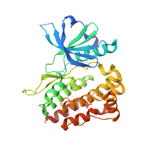
4L00

Crystal structure of the apo Jak1 pseudokinase domain


Experimental Data Snapshot

  • Method: X-RAY DIFFRACTION
  • Resolution: 1.80 Å
  • R-Value Free: 0.203 
  • R-Value Work: 0.169 
  • R-Value Observed: 0.171 

wwPDB Validation   3D Report Full Report


This is version 1.3 of the entry. See complete history


Literature

Structure of a pseudokinase-domain switch that controls oncogenic activation of Jak kinases.

Toms, A.V.Deshpande, A.McNally, R.Jeong, Y.Rogers, J.M.Kim, C.U.Gruner, S.M.Ficarro, S.B.Marto, J.A.Sattler, M.Griffin, J.D.Eck, M.J.

(2013) Nat Struct Mol Biol 20: 1221-1223

  • DOI: https://doi.org/10.1038/nsmb.2673
  • Primary Citation of Related Structures:  
    4L00, 4L01

  • PubMed Abstract: 

    The V617F mutation in the Jak2 pseudokinase domain causes myeloproliferative neoplasms, and the equivalent mutation in Jak1 (V658F) is found in T-cell leukemias. Crystal structures of wild-type and V658F-mutant human Jak1 pseudokinase reveal a conformational switch that remodels a linker segment encoded by exon 12, which is also a site of mutations in Jak2. This switch is required for V617F-mediated Jak2 activation and possibly for physiologic Jak activation.


  • Organizational Affiliation

    1] Department of Biological Chemistry and Molecular Pharmacology, Harvard Medical School, Boston, Massachusetts, USA. [2] Department of Cancer Biology, Dana-Farber Cancer Institute, Boston, Massachusetts, USA.


Macromolecules
Find similar proteins by:  (by identity cutoff)  |  3D Structure
Entity ID: 1
MoleculeChains Sequence LengthOrganismDetailsImage
Tyrosine-protein kinase JAK1
A, B
304Homo sapiensMutation(s): 0 
Gene Names: JAK1JAK1AJAK1B
EC: 2.7.10.2
UniProt & NIH Common Fund Data Resources
Find proteins for P23458 (Homo sapiens)
Explore P23458 
Go to UniProtKB:  P23458
PHAROS:  P23458
GTEx:  ENSG00000162434 
Entity Groups  
Sequence Clusters30% Identity50% Identity70% Identity90% Identity95% Identity100% Identity
UniProt GroupP23458
Sequence Annotations
Expand
  • Reference Sequence
Experimental Data & Validation

Experimental Data

  • Method: X-RAY DIFFRACTION
  • Resolution: 1.80 Å
  • R-Value Free: 0.203 
  • R-Value Work: 0.169 
  • R-Value Observed: 0.171 
  • Space Group: P 1 21 1
Unit Cell:
Length ( Å )Angle ( ˚ )
a = 47.026α = 90
b = 80.656β = 95.44
c = 76.231γ = 90
Software Package:
Software NamePurpose
PHASERphasing
REFMACrefinement
HKL-2000data reduction
HKL-2000data scaling

Structure Validation

View Full Validation Report


Entry History 

Deposition Data

Revision History  (Full details and data files)

  • Version 1.0: 2013-09-04
    Type: Initial release
  • Version 1.1: 2013-10-02
    Changes: Database references
  • Version 1.2: 2014-02-05
    Changes: Database references
  • Version 1.3: 2024-02-28
    Changes: Data collection, Database references