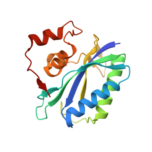
4M5L

The Identification, Analysis and Structure-Based Development of Novel Inhibitors of 6-hydroxymethyl-7,8-dihydropterin pyrophosphokinase


Experimental Data Snapshot

  • Method: X-RAY DIFFRACTION
  • Resolution: 1.09 Å
  • R-Value Free: 0.186 
  • R-Value Work: 0.173 
  • R-Value Observed: 0.173 

Starting Model: experimental
View more details

wwPDB Validation   3D Report Full Report


Ligand Structure Quality Assessment 


This is version 1.2 of the entry. See complete history


Literature

The identification, analysis and structure-based development of novel inhibitors of 6-hydroxymethyl-7,8-dihydropterin pyrophosphokinase.

Yun, M.K.Hoagland, D.Kumar, G.Waddell, M.B.Rock, C.O.Lee, R.E.White, S.W.

(2014) Bioorg Med Chem 22: 2157-2165

  • DOI: https://doi.org/10.1016/j.bmc.2014.02.022
  • Primary Citation of Related Structures:  
    4M5G, 4M5H, 4M5I, 4M5J, 4M5K, 4M5L, 4M5M, 4M5N

  • PubMed Abstract: 

    6-Hydroxymethyl-7,8-dihydropterin pyrophosphokinase (HPPK) is an essential enzyme in the microbial folate biosynthetic pathway. This pathway has proven to be an excellent target for antimicrobial development, but widespread resistance to common therapeutics including the sulfa drugs has stimulated interest in HPPK as an alternative target in the pathway. A screen of a pterin-biased compound set identified several HPPK inhibitors that contain an aryl substituted 8-thioguanine scaffold, and structural analyses showed that these compounds engage the HPPK pterin-binding pocket and an induced cryptic pocket. A preliminary structure activity relationship profile was developed from biophysical and biochemical characterizations of derivative molecules. Also, a similarity search identified additional scaffolds that bind more tightly within the HPPK pterin pocket. These inhibitory scaffolds have the potential for rapid elaboration into novel lead antimicrobial agents.


  • Organizational Affiliation

    Department of Structural Biology, St. Jude Children's Research Hospital, Memphis, TN 38105, USA.


Macromolecules
Find similar proteins by:  (by identity cutoff)  |  3D Structure
Entity ID: 1
MoleculeChains Sequence LengthOrganismDetailsImage
2-amino-4-hydroxy-6-hydroxymethyldihydropteridine pyrophosphokinase162Escherichia coli K-12Mutation(s): 0 
Gene Names: folKb0142JW0138
EC: 2.7.6.3
UniProt
Find proteins for P26281 (Escherichia coli (strain K12))
Explore P26281 
Go to UniProtKB:  P26281
Entity Groups  
Sequence Clusters30% Identity50% Identity70% Identity90% Identity95% Identity100% Identity
UniProt GroupP26281
Sequence Annotations
Expand
  • Reference Sequence
Small Molecules
Ligands 4 Unique
IDChains Name / Formula / InChI Key2D Diagram3D Interactions
APC
Query on APC

Download Ideal Coordinates CCD File 
B [auth A]DIPHOSPHOMETHYLPHOSPHONIC ACID ADENOSYL ESTER
C11 H18 N5 O12 P3
CAWZRIXWFRFUQB-IOSLPCCCSA-N
YH4
Query on YH4

Download Ideal Coordinates CCD File 
G [auth A]2-amino-8-{[2-(2-methylphenyl)-2-oxoethyl]sulfanyl}-1,7-dihydro-6H-purin-6-one
C14 H13 N5 O2 S
YXZULINHSYINAQ-UHFFFAOYSA-N
GOL
Query on GOL

Download Ideal Coordinates CCD File 
F [auth A]GLYCEROL
C3 H8 O3
PEDCQBHIVMGVHV-UHFFFAOYSA-N
CA
Query on CA

Download Ideal Coordinates CCD File 
C [auth A],
D [auth A],
E [auth A]
CALCIUM ION
Ca
BHPQYMZQTOCNFJ-UHFFFAOYSA-N
Experimental Data & Validation

Experimental Data

  • Method: X-RAY DIFFRACTION
  • Resolution: 1.09 Å
  • R-Value Free: 0.186 
  • R-Value Work: 0.173 
  • R-Value Observed: 0.173 
  • Space Group: P 1 21 1
Unit Cell:
Length ( Å )Angle ( ˚ )
a = 35.903α = 90
b = 57.991β = 115.49
c = 38.643γ = 90
Software Package:
Software NamePurpose
SERGUIdata collection
PHENIXmodel building
PHENIXrefinement
HKL-2000data reduction
HKL-2000data scaling
PHENIXphasing

Structure Validation

View Full Validation Report


Ligand Structure Quality Assessment 


Entry History 

Deposition Data

Revision History  (Full details and data files)

  • Version 1.0: 2014-04-02
    Type: Initial release
  • Version 1.1: 2014-04-16
    Changes: Database references
  • Version 1.2: 2023-09-20
    Changes: Data collection, Database references, Derived calculations, Refinement description