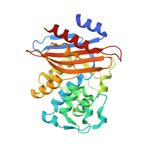
Perturbing the General Base Residue Glu166 in the Active Site of Class A beta-Lactamase Leads to Enhanced Carbapenem Binding and Acylation
Pan, X., Wong, W., He, Y., Jiang, Y., Zhao, Y.(2014) Biochemistry 53: 5414-5423
- PubMed: 25020031
- DOI: https://doi.org/10.1021/bi401609h
- Primary Citation of Related Structures:
4N92, 4N9K, 4N9L - PubMed Abstract:
Most class A β-lactamases cannot hydrolyze carbapenem antibiotics effectively. The molecular mechanism of this catalytic inefficiency has been attributed to the unique stereochemistry of carbapenems, including their 6-α-hydroxyethyl side chain and the transition between two tautomeric states when bound at the active site. Previous studies have shown that the 6-α-hydroxyethyl side chain of carbapenems can interfere with catalysis by forming hydrogen bonds with the deacylation water molecule to reduce its nucleophilicity. Here our studies of a class A noncarbapenemase PenP demonstrate that substituting the general base residue Glu166 with Ser or other residues leads to a significant enhancement of the acylation kinetics by ∼100-500 times toward carbapenems like meropenem. The structures of PenP and Glu166Ser both in apo form and in complex with meropenem reveal that Glu166 is critical for the formation of a hydrogen bonding network within the active site that locks Asn170 in an orientation to impose steric clash with the 6-α-hydroxyethyl side chain of meropenem. The Glu166Ser substitution weakens this network and enables Asn170 to adopt an alternative conformation to avoid steric clash and accommodate faster acylation kinetics. Furthermore, the weakened hydrogen bonding network caused by the Glu166Ser substitution allows the 6-α-hydroxyethyl moiety to adopt a catalytically favorable orientation as seen in class A carbapenemases. In summary, our data identify a previously unreported role of the universally conserved general base residue Glu166 in impeding the proper binding of carbapenems by restricting their 6-α-hydroxyethyl group.
Organizational Affiliation:
Department of Applied Biology and Chemical Technology, State Key Laboratory of Chirosciences, The Hong Kong Polytechnic University , Hung Hom, Kowloon, Hong Kong, P. R. China.


