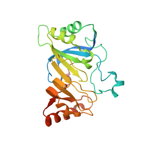
4NJH

Crystal Structure of QueE from Burkholderia multivorans in complex with AdoMet and 6-carboxy-5,6,7,8-tetrahydropterin


Experimental Data Snapshot

  • Method: X-RAY DIFFRACTION
  • Resolution: 1.90 Å
  • R-Value Free: 0.174 
  • R-Value Work: 0.147 
  • R-Value Observed: 0.148 

Starting Model: experimental
View more details

wwPDB Validation   3D Report Full Report


Ligand Structure Quality Assessment 


This is version 1.4 of the entry. See complete history


Literature

Radical SAM enzyme QueE defines a new minimal core fold and metal-dependent mechanism.

Dowling, D.P.Bruender, N.A.Young, A.P.McCarty, R.M.Bandarian, V.Drennan, C.L.

(2014) Nat Chem Biol 10: 106-112

  • DOI: https://doi.org/10.1038/nchembio.1426
  • Primary Citation of Related Structures:  
    4NJG, 4NJH, 4NJI, 4NJJ, 4NJK

  • PubMed Abstract: 

    7-carboxy-7-deazaguanine synthase (QueE) catalyzes a key S-adenosyl-L-methionine (AdoMet)- and Mg(2+)-dependent radical-mediated ring contraction step, which is common to the biosynthetic pathways of all deazapurine-containing compounds. QueE is a member of the AdoMet radical superfamily, which employs the 5'-deoxyadenosyl radical from reductive cleavage of AdoMet to initiate chemistry. To provide a mechanistic rationale for this elaborate transformation, we present the crystal structure of a QueE along with structures of pre- and post-turnover states. We find that substrate binds perpendicular to the [4Fe-4S]-bound AdoMet, exposing its C6 hydrogen atom for abstraction and generating the binding site for Mg(2+), which coordinates directly to the substrate. The Burkholderia multivorans structure reported here varies from all other previously characterized members of the AdoMet radical superfamily in that it contains a hypermodified (β6/α3) protein core and an expanded cluster-binding motif, CX14CX2C.


  • Organizational Affiliation

    1] Howard Hughes Medical Institute, Massachusetts Institute of Technology, Cambridge, Massachusetts, USA. [2] Department of Chemistry, Massachusetts Institute of Technology, Cambridge, Massachusetts, USA.


Macromolecules
Find similar proteins by:  (by identity cutoff)  |  3D Structure
Entity ID: 1
MoleculeChains Sequence LengthOrganismDetailsImage
7-carboxy-7-deazaguanine synthase
A, B
230Burkholderia multivorans ATCC 17616Mutation(s): 0 
Gene Names: queEBmul_3115BMULJ_00116
EC: 4.3.99.3
UniProt
Find proteins for A0A0H3KB22 (Burkholderia multivorans (strain ATCC 17616 / 249))
Explore A0A0H3KB22 
Go to UniProtKB:  A0A0H3KB22
Entity Groups  
Sequence Clusters30% Identity50% Identity70% Identity90% Identity95% Identity100% Identity
UniProt GroupA0A0H3KB22
Sequence Annotations
Expand
  • Reference Sequence
Small Molecules
Ligands 5 Unique
IDChains Name / Formula / InChI Key2D Diagram3D Interactions
SAM
Query on SAM

Download Ideal Coordinates CCD File 
D [auth A],
I [auth B]
S-ADENOSYLMETHIONINE
C15 H22 N6 O5 S
MEFKEPWMEQBLKI-FCKMPRQPSA-N
SF4
Query on SF4

Download Ideal Coordinates CCD File 
C [auth A],
H [auth B]
IRON/SULFUR CLUSTER
Fe4 S4
LJBDFODJNLIPKO-UHFFFAOYSA-N
2K8
Query on 2K8

Download Ideal Coordinates CCD File 
E [auth A],
J [auth B]
(6R)-2-amino-4-oxo-3,4,5,6,7,8-hexahydropteridine-6-carboxylic acid
C7 H9 N5 O3
QSIYONWVWDSRRO-UWTATZPHSA-N
PO4
Query on PO4

Download Ideal Coordinates CCD File 
G [auth A],
L [auth B]
PHOSPHATE ION
O4 P
NBIIXXVUZAFLBC-UHFFFAOYSA-K
NA
Query on NA

Download Ideal Coordinates CCD File 
F [auth A],
K [auth B],
M [auth B]
SODIUM ION
Na
FKNQFGJONOIPTF-UHFFFAOYSA-N
Experimental Data & Validation

Experimental Data

  • Method: X-RAY DIFFRACTION
  • Resolution: 1.90 Å
  • R-Value Free: 0.174 
  • R-Value Work: 0.147 
  • R-Value Observed: 0.148 
  • Space Group: P 43 21 2
Unit Cell:
Length ( Å )Angle ( ˚ )
a = 119.129α = 90
b = 119.129β = 90
c = 103.363γ = 90
Software Package:
Software NamePurpose
ADSCdata collection
PHENIXmodel building
PHENIXrefinement
HKL-2000data reduction
HKL-2000data scaling
PHENIXphasing

Structure Validation

View Full Validation Report


Ligand Structure Quality Assessment 


Entry History 

Deposition Data

Revision History  (Full details and data files)

  • Version 1.0: 2013-12-25
    Type: Initial release
  • Version 1.1: 2014-01-22
    Changes: Database references
  • Version 1.2: 2014-02-05
    Changes: Database references
  • Version 1.3: 2014-07-02
    Changes: Non-polymer description
  • Version 1.4: 2023-09-20
    Changes: Data collection, Database references, Derived calculations, Refinement description