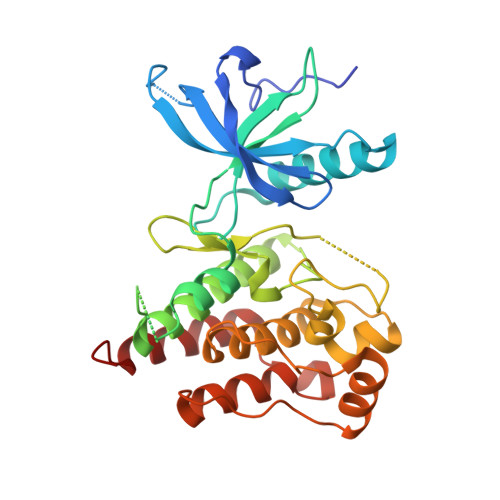
4NK9

Crystal structure of human fibroblast growth factor receptor 1 kinase domain in complex with pyrazolaminopyrimidine 1


Experimental Data Snapshot

  • Method: X-RAY DIFFRACTION
  • Resolution: 2.57 Å
  • R-Value Free: 0.262 
  • R-Value Work: 0.206 
  • R-Value Observed: 0.209 

wwPDB Validation   3D Report Full Report


Ligand Structure Quality Assessment 


This is version 1.2 of the entry. See complete history


Literature

FGFR1 Kinase Inhibitors: Close Regioisomers Adopt Divergent Binding Modes and Display Distinct Biophysical Signatures.

Klein, T.Tucker, J.Holdgate, G.A.Norman, R.A.Breeze, A.L.

(2014) ACS Med Chem Lett 5: 166-171

  • DOI: https://doi.org/10.1021/ml4004205
  • Primary Citation of Related Structures:  
    4NK9, 4NKA, 4NKS

  • PubMed Abstract: 

    The binding of a ligand to its target protein is often accompanied by conformational changes of both the protein and the ligand. This is of particular interest, since structural rearrangements of the macromolecular target and the ligand influence the free energy change upon complex formation. In this study, we use X-ray crystallography, isothermal titration calorimetry, and surface-plasmon resonance biosensor analysis to investigate the binding of pyrazolylaminopyrimidine inhibitors to FGFR1 tyrosine kinase, an important anticancer target. Our results highlight that structurally close analogs of this inhibitor series interact with FGFR1 with different binding modes, which are a consequence of conformational changes in both the protein and the ligand as well as the bound water network. Together with the collected kinetic and thermodynamic data, we use the protein-ligand crystal structure information to rationalize the observed inhibitory potencies on a molecular level.


  • Organizational Affiliation

    Discovery Sciences, AstraZeneca R&D , Alderley Park, Macclesfield, Cheshire, SK10 4TG, U.K.


Macromolecules
Find similar proteins by:  (by identity cutoff)  |  3D Structure
Entity ID: 1
MoleculeChains Sequence LengthOrganismDetailsImage
FIBROBLAST GROWTH FACTOR RECEPTOR 1
A, B
309Homo sapiensMutation(s): 2 
Gene Names: FGFR1BFGFRCEKFGFBRFLGFLT2HBGFR
EC: 2.7.10.1
UniProt & NIH Common Fund Data Resources
Find proteins for P11362 (Homo sapiens)
Explore P11362 
Go to UniProtKB:  P11362
PHAROS:  P11362
GTEx:  ENSG00000077782 
Entity Groups  
Sequence Clusters30% Identity50% Identity70% Identity90% Identity95% Identity100% Identity
UniProt GroupP11362
Sequence Annotations
Expand
  • Reference Sequence
Small Molecules
Ligands 3 Unique
IDChains Name / Formula / InChI Key2D Diagram3D Interactions
2K5
Query on 2K5

Download Ideal Coordinates CCD File 
I [auth A],
K [auth B]
N~4~-{5-[2-(3,5-dimethoxyphenyl)ethyl]-1H-pyrazol-3-yl}-N~2~-[(3-methyl-1,2-oxazol-5-yl)methyl]pyrimidine-2,4-diamine
C22 H25 N7 O3
AQHXGQTWGFVXTB-UHFFFAOYSA-N
SO4
Query on SO4

Download Ideal Coordinates CCD File 
G [auth A],
H [auth A],
J [auth B]
SULFATE ION
O4 S
QAOWNCQODCNURD-UHFFFAOYSA-L
EDO
Query on EDO

Download Ideal Coordinates CCD File 
C [auth A],
D [auth A],
E [auth A],
F [auth A]
1,2-ETHANEDIOL
C2 H6 O2
LYCAIKOWRPUZTN-UHFFFAOYSA-N
Binding Affinity Annotations 
IDSourceBinding Affinity
2K5 PDBBind:  4NK9 Kd: 3.1 (nM) from 1 assay(s)
Experimental Data & Validation

Experimental Data

  • Method: X-RAY DIFFRACTION
  • Resolution: 2.57 Å
  • R-Value Free: 0.262 
  • R-Value Work: 0.206 
  • R-Value Observed: 0.209 
  • Space Group: C 1 2 1
Unit Cell:
Length ( Å )Angle ( ˚ )
a = 210.34α = 90
b = 55.97β = 107.43
c = 65.72γ = 90
Software Package:
Software NamePurpose
GDAdata collection
AMoREphasing
BUSTERrefinement
XDSdata reduction
SCALAdata scaling

Structure Validation

View Full Validation Report


Ligand Structure Quality Assessment 


Entry History 

Deposition Data

Revision History  (Full details and data files)

  • Version 1.0: 2013-12-18
    Type: Initial release
  • Version 1.1: 2014-07-16
    Changes: Database references
  • Version 1.2: 2024-02-28
    Changes: Data collection, Database references, Derived calculations