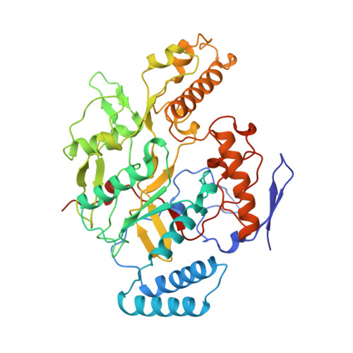
4NOS

HUMAN INDUCIBLE NITRIC OXIDE SYNTHASE WITH INHIBITOR


Experimental Data Snapshot

  • Method: X-RAY DIFFRACTION
  • Resolution: 2.25 Å
  • R-Value Free: 0.289 
  • R-Value Work: 0.199 

wwPDB Validation   3D Report Full Report


This is version 1.3 of the entry. See complete history


Literature

Structural characterization of nitric oxide synthase isoforms reveals striking active-site conservation.

Fischmann, T.O.Hruza, A.Niu, X.D.Fossetta, J.D.Lunn, C.A.Dolphin, E.Prongay, A.J.Reichert, P.Lundell, D.J.Narula, S.K.Weber, P.C.

(1999) Nat Struct Biol 6: 233-242

  • DOI: https://doi.org/10.1038/6675
  • Primary Citation of Related Structures:  
    3NOS, 4NOS

  • PubMed Abstract: 

    Crystal structures of human endothelial nitric oxide synthase (eNOS) and human inducible NOS (iNOS) catalytic domains were solved in complex with the arginine substrate and an inhibitor S-ethylisothiourea (SEITU), respectively. The small molecules bind in a narrow cleft within the larger active-site cavity containing heme and tetrahydrobiopterin. Both are hydrogen-bonded to a conserved glutamate (eNOS E361, iNOS E377). The active-site residues of iNOS and eNOS are nearly identical. Nevertheless, structural comparisons provide a basis for design of isozyme-selective inhibitors. The high-resolution, refined structures of eNOS (2.4 A resolution) and iNOS (2.25 A resolution) reveal an unexpected structural zinc situated at the intermolecular interface and coordinated by four cysteines, two from each monomer.


  • Organizational Affiliation

    Structural Chemistry Department, Schering-Plough Research Institute, Kenilworth, New Jersey 07033, USA. [email protected]


Macromolecules
Find similar proteins by:  (by identity cutoff)  |  3D Structure
Entity ID: 1
MoleculeChains Sequence LengthOrganismDetailsImage
INDUCIBLE NITRIC OXIDE SYNTHASE
A, B, C, D
427Homo sapiensMutation(s): 0 
EC: 1.14.13.39
UniProt & NIH Common Fund Data Resources
Find proteins for P35228 (Homo sapiens)
Explore P35228 
Go to UniProtKB:  P35228
PHAROS:  P35228
GTEx:  ENSG00000007171 
Entity Groups  
Sequence Clusters30% Identity50% Identity70% Identity90% Identity95% Identity100% Identity
UniProt GroupP35228
Sequence Annotations
Expand
  • Reference Sequence
Small Molecules
Ligands 5 Unique
IDChains Name / Formula / InChI Key2D Diagram3D Interactions
HEM
Query on HEM

Download Ideal Coordinates CCD File 
F [auth A],
I [auth B],
M [auth C],
P [auth D]
PROTOPORPHYRIN IX CONTAINING FE
C34 H32 Fe N4 O4
KABFMIBPWCXCRK-RGGAHWMASA-L
H4B
Query on H4B

Download Ideal Coordinates CCD File 
J [auth B],
N [auth C],
Q [auth D]
5,6,7,8-TETRAHYDROBIOPTERIN
C9 H15 N5 O3
FNKQXYHWGSIFBK-RPDRRWSUSA-N
H2B
Query on H2B

Download Ideal Coordinates CCD File 
G [auth A]2-AMINO-6-(1,2-DIHYDROXY-PROPYL)-7,8-DIHYDRO-6H-PTERIDIN-4-ONE
C9 H13 N5 O3
ZHQJVZLJDXWFFX-RPDRRWSUSA-N
ITU
Query on ITU

Download Ideal Coordinates CCD File 
H [auth A],
K [auth B],
O [auth C],
R [auth D]
ETHYLISOTHIOUREA
C3 H8 N2 S
VFIZBHJTOHUOEK-UHFFFAOYSA-N
ZN
Query on ZN

Download Ideal Coordinates CCD File 
E [auth A],
L [auth C]
ZINC ION
Zn
PTFCDOFLOPIGGS-UHFFFAOYSA-N
Experimental Data & Validation

Experimental Data

  • Method: X-RAY DIFFRACTION
  • Resolution: 2.25 Å
  • R-Value Free: 0.289 
  • R-Value Work: 0.199 
  • Space Group: P 21 21 21
Unit Cell:
Length ( Å )Angle ( ˚ )
a = 89.759α = 90
b = 156.672β = 90
c = 190.044γ = 90
Software Package:
Software NamePurpose
SHARPphasing
SOLOMONphasing
X-PLORrefinement
DENZOdata reduction
CCP4data scaling

Structure Validation

View Full Validation Report


Entry History 

Deposition Data

Revision History  (Full details and data files)

  • Version 1.0: 2000-02-04
    Type: Initial release
  • Version 1.1: 2007-10-16
    Changes: Version format compliance
  • Version 1.2: 2011-07-13
    Changes: Derived calculations, Version format compliance
  • Version 1.3: 2023-12-27
    Changes: Data collection, Database references, Derived calculations