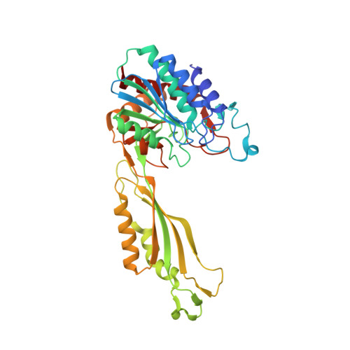
Inhibition of the dapE-Encoded N-Succinyl-L,L-diaminopimelic Acid Desuccinylase from Neisseria meningitidis by L-Captopril.
Starus, A., Nocek, B., Bennett, B., Larrabee, J.A., Shaw, D.L., Sae-Lee, W., Russo, M.T., Gillner, D.M., Makowska-Grzyska, M., Joachimiak, A., Holz, R.C.(2015) Biochemistry 54: 4834-4844
- PubMed: 26186504
- DOI: https://doi.org/10.1021/acs.biochem.5b00475
- Primary Citation of Related Structures:
4O23, 4PPZ, 4PQA - PubMed Abstract:
Binding of the competitive inhibitor L-captopril to the dapE-encoded N-succinyl-L,L-diaminopimelic acid desuccinylase from Neisseria meningitidis (NmDapE) was examined by kinetic, spectroscopic, and crystallographic methods. L-Captopril, an angiotensin-converting enzyme (ACE) inhibitor, was previously shown to be a potent inhibitor of the DapE from Haemophilus influenzae (HiDapE) with an IC50 of 3.3 μM and a measured Ki of 1.8 μM and displayed a dose-responsive antibiotic activity toward Escherichia coli. L-Captopril is also a competitive inhibitor of NmDapE with a Ki of 2.8 μM. To examine the nature of the interaction of L-captopril with the dinuclear active site of DapE, we have obtained electron paramagnetic resonance (EPR) and magnetic circular dichroism (MCD) data for the enzymatically hyperactive Co(II)-substituted forms of both HiDapE and NmDapE. EPR and MCD data indicate that the two Co(II) ions in DapE are antiferromagnetically coupled, yielding an S = 0 ground state, and suggest a thiolate bridge between the two metal ions. Verification of a thiolate-bridged dinuclear complex was obtained by determining the three-dimensional X-ray crystal structure of NmDapE in complex with L-captopril at 1.8 Å resolution. Combination of these data provides new insights into binding of L-captopril to the active site of DapE enzymes as well as important inhibitor-active site residue interaction's. Such information is critical for the design of new, potent inhibitors of DapE enzymes.
Organizational Affiliation:
†Department of Chemistry and Biochemistry, Loyola University-Chicago, 1068 West Sheridan Road, Chicago, Illinois 60626, United States.


