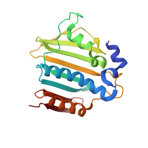
4P8O

S. aureus gyrase bound to an aminobenzimidazole urea inhibitor


Experimental Data Snapshot

  • Method: X-RAY DIFFRACTION
  • Resolution: 2.40 Å
  • R-Value Free: 0.239 
  • R-Value Work: 0.188 
  • R-Value Observed: 0.191 

wwPDB Validation   3D Report Full Report


Ligand Structure Quality Assessment 


This is version 1.3 of the entry. See complete history


Literature

Second-generation antibacterial benzimidazole ureas: discovery of a preclinical candidate with reduced metabolic liability.

Grillot, A.L.Tiran, A.L.Shannon, D.Krueger, E.Liao, Y.O'Dowd, H.Tang, Q.Ronkin, S.Wang, T.Waal, N.Li, P.Lauffer, D.Sizensky, E.Tanoury, J.Perola, E.Grossman, T.H.Doyle, T.Hanzelka, B.Jones, S.Dixit, V.Ewing, N.Liao, S.Boucher, B.Jacobs, M.Bennani, Y.Charifson, P.S.

(2014) J Med Chem 57: 8792-8816

  • DOI: https://doi.org/10.1021/jm500563g
  • Primary Citation of Related Structures:  
    4P8O

  • PubMed Abstract: 

    Compound 3 is a potent aminobenzimidazole urea with broad-spectrum Gram-positive antibacterial activity resulting from dual inhibition of bacterial gyrase (GyrB) and topoisomerase IV (ParE), and it demonstrates efficacy in rodent models of bacterial infection. Preclinical in vitro and in vivo studies showed that compound 3 covalently labels liver proteins, presumably via formation of a reactive metabolite, and hence presented a potential safety liability. The urea moiety in compound 3 was identified as being potentially responsible for reactive metabolite formation, but its replacement resulted in loss of antibacterial activity and/or oral exposure due to poor physicochemical parameters. To identify second-generation aminobenzimidazole ureas devoid of reactive metabolite formation potential, we implemented a metabolic shift strategy, which focused on shifting metabolism away from the urea moiety by introducing metabolic soft spots elsewhere in the molecule. Aminobenzimidazole urea 34, identified through this strategy, exhibits similar antibacterial activity as that of 3 and did not label liver proteins in vivo, indicating reduced/no potential for reactive metabolite formation.


  • Organizational Affiliation

    Vertex Pharmaceuticals Incorporated , 50 Northern Avenue, Boston, Massachusetts 02210, United States.


Macromolecules
Find similar proteins by:  (by identity cutoff)  |  3D Structure
Entity ID: 1
MoleculeChains Sequence LengthOrganismDetailsImage
DNA gyrase subunit B
A, B
187Staphylococcus aureus 04-02981Mutation(s): 0 
Gene Names: gyrBSAR0005
EC: 5.99.1.3 (PDB Primary Data), 5.6.2.2 (UniProt)
UniProt
Find proteins for Q6GKU0 (Staphylococcus aureus (strain MRSA252))
Explore Q6GKU0 
Go to UniProtKB:  Q6GKU0
Entity Groups  
Sequence Clusters30% Identity50% Identity70% Identity90% Identity95% Identity100% Identity
UniProt GroupQ6GKU0
Sequence Annotations
Expand
  • Reference Sequence
Small Molecules
Ligands 1 Unique
IDChains Name / Formula / InChI Key2D Diagram3D Interactions
883
Query on 883

Download Ideal Coordinates CCD File 
C [auth A],
D [auth B]
1-ethyl-3-[5-(5-fluoropyridin-3-yl)-7-(pyrimidin-2-yl)-1H-benzimidazol-2-yl]urea
C19 H16 F N7 O
FKCGKEHVIVGGDU-UHFFFAOYSA-N
Experimental Data & Validation

Experimental Data

  • Method: X-RAY DIFFRACTION
  • Resolution: 2.40 Å
  • R-Value Free: 0.239 
  • R-Value Work: 0.188 
  • R-Value Observed: 0.191 
  • Space Group: C 1 2 1
Unit Cell:
Length ( Å )Angle ( ˚ )
a = 143.46α = 90
b = 55.73β = 100.42
c = 50.8γ = 90
Software Package:
Software NamePurpose
SCALEPACKdata scaling
BUSTER-TNTrefinement
PDB_EXTRACTdata extraction

Structure Validation

View Full Validation Report


Ligand Structure Quality Assessment 


Entry History 

Deposition Data

  • Released Date: 2014-10-29 
  • Deposition Author(s): Jacobs, M.D.

Revision History  (Full details and data files)

  • Version 1.0: 2014-10-29
    Type: Initial release
  • Version 1.1: 2014-11-26
    Changes: Database references
  • Version 1.2: 2015-09-30
    Changes: Data collection
  • Version 1.3: 2023-12-27
    Changes: Data collection, Database references, Derived calculations