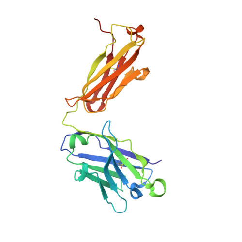
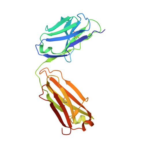
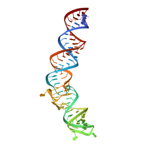
4Q9Q

Crystal structure of an RNA aptamer bound to bromo-ligand analog in complex with Fab


Experimental Data Snapshot

  • Method: X-RAY DIFFRACTION
  • Resolution: 2.45 Å
  • R-Value Free: 0.221 
  • R-Value Work: 0.182 
  • R-Value Observed: 0.184 

wwPDB Validation   3D Report Full Report


Ligand Structure Quality Assessment 


This is version 2.1 of the entry. See complete history


Literature

A G-quadruplex-containing RNA activates fluorescence in a GFP-like fluorophore.

Huang, H.Suslov, N.B.Li, N.S.Shelke, S.A.Evans, M.E.Koldobskaya, Y.Rice, P.A.Piccirilli, J.A.

(2014) Nat Chem Biol 10: 686-691

  • DOI: https://doi.org/10.1038/nchembio.1561
  • Primary Citation of Related Structures:  
    4KZD, 4KZE, 4Q9Q, 4Q9R

  • PubMed Abstract: 

    Spinach is an in vitro-selected RNA aptamer that binds a GFP-like ligand and activates its green fluorescence. Spinach is thus an RNA analog of GFP and has potentially widespread applications for in vivo labeling and imaging. We used antibody-assisted crystallography to determine the structures of Spinach both with and without bound fluorophore at 2.2-Å and 2.4-Å resolution, respectively. Spinach RNA has an elongated structure containing two helical domains separated by an internal bulge that folds into a G-quadruplex motif of unusual topology. The G-quadruplex motif and adjacent nucleotides comprise a partially preformed binding site for the fluorophore. The fluorophore binds in a planar conformation and makes extensive aromatic stacking and hydrogen bond interactions with the RNA. Our findings provide a foundation for structure-based engineering of new fluorophore-binding RNA aptamers.


  • Organizational Affiliation

    Department of Chemistry, University of Chicago, Chicago, Illinois, USA.


Macromolecules

Find similar proteins by:  (by identity cutoff)  |  3D Structure
Entity ID: 1
MoleculeChains Sequence LengthOrganismDetailsImage
Fab BL3-6, HEAVY CHAINA [auth H]225Mus musculusMutation(s): 0 
Entity Groups  
Sequence Clusters30% Identity50% Identity70% Identity90% Identity95% Identity100% Identity
Sequence Annotations
Expand
  • Reference Sequence
Find similar proteins by:  (by identity cutoff)  |  3D Structure
Entity ID: 2
MoleculeChains Sequence LengthOrganismDetailsImage
Fab BL3-6, LIGHT CHAINB [auth L]215Mus musculusMutation(s): 0 
Entity Groups  
Sequence Clusters30% Identity50% Identity70% Identity90% Identity95% Identity100% Identity
Sequence Annotations
Expand
  • Reference Sequence
Find similar nucleic acids by:  (by identity cutoff)  |  3D Structure
Entity ID: 3
MoleculeChains LengthOrganismImage
Spinach RNA aptamerC [auth R]84synthetic construct
Sequence Annotations
Expand
  • Reference Sequence
Experimental Data & Validation

Experimental Data

  • Method: X-RAY DIFFRACTION
  • Resolution: 2.45 Å
  • R-Value Free: 0.221 
  • R-Value Work: 0.182 
  • R-Value Observed: 0.184 
  • Space Group: C 1 2 1
Unit Cell:
Length ( Å )Angle ( ˚ )
a = 146.545α = 90
b = 79.676β = 111.51
c = 95.28γ = 90
Software Package:
Software NamePurpose
Blu-Icedata collection
PHENIXmodel building
PHENIXrefinement
HKL-2000data reduction
HKL-2000data scaling
PHENIXphasing

Structure Validation

View Full Validation Report


Ligand Structure Quality Assessment 


Entry History 

Deposition Data

Revision History  (Full details and data files)

  • Version 1.0: 2014-06-18
    Type: Initial release
  • Version 1.1: 2014-07-09
    Changes: Database references
  • Version 1.2: 2014-07-30
    Changes: Database references
  • Version 1.3: 2014-10-15
    Changes: Structure summary
  • Version 2.0: 2017-07-26
    Changes: Advisory, Atomic model, Database references, Derived calculations, Other, Polymer sequence, Source and taxonomy, Structure summary
  • Version 2.1: 2024-11-06
    Changes: Data collection, Database references, Derived calculations, Structure summary