4QZG

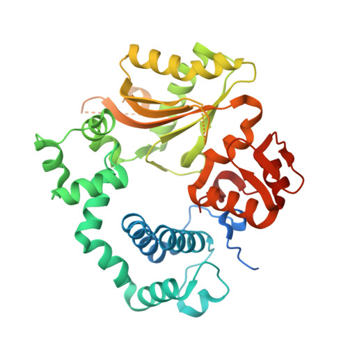
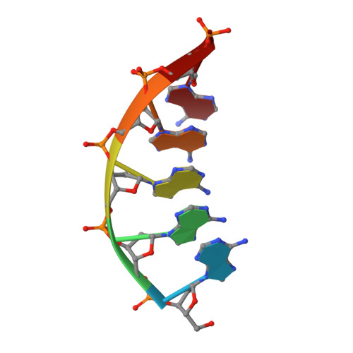
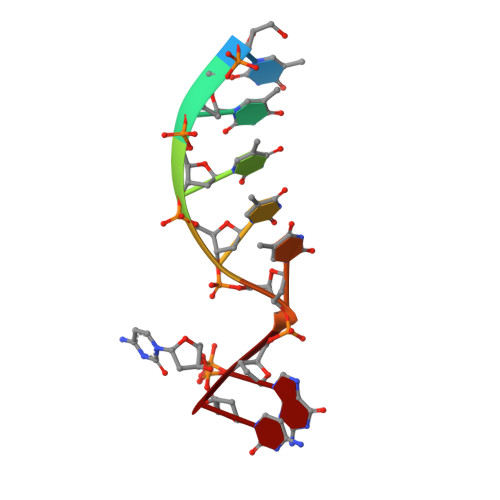
Mouse Tdt, F401A mutant, in complex with a DSB substrate, C-C base pair


Experimental Data Snapshot

  • Method: X-RAY DIFFRACTION
  • Resolution: 2.75 Å
  • R-Value Free: 0.208 
  • R-Value Work: 0.188 
  • R-Value Observed: 0.189 

Starting Model: experimental
View more details

wwPDB Validation   3D Report Full Report


Ligand Structure Quality Assessment 


This is version 1.2 of the entry. See complete history


Literature

Structural basis for a novel mechanism of DNA bridging and alignment in eukaryotic DSB DNA repair.

Gouge, J.Rosario, S.Romain, F.Poitevin, F.Beguin, P.Delarue, M.

(2015) EMBO J 34: 1126-1142

  • DOI: https://doi.org/10.15252/embj.201489643
  • Primary Citation of Related Structures:  
    4QZ8, 4QZ9, 4QZA, 4QZB, 4QZC, 4QZD, 4QZE, 4QZF, 4QZG, 4QZH, 4QZI

  • PubMed Abstract: 

    Eukaryotic DNA polymerase mu of the PolX family can promote the association of the two 3'-protruding ends of a DNA double-strand break (DSB) being repaired (DNA synapsis) even in the absence of the core non-homologous end-joining (NHEJ) machinery. Here, we show that terminal deoxynucleotidyltransferase (TdT), a closely related PolX involved in V(D)J recombination, has the same property. We solved its crystal structure with an annealed DNA synapsis containing one micro-homology (MH) base pair and one nascent base pair. This structure reveals how the N-terminal domain and Loop 1 of Tdt cooperate for bridging the two DNA ends, providing a templating base in trans and limiting the MH search region to only two base pairs. A network of ordered water molecules is proposed to assist the incorporation of any nucleotide independently of the in trans templating base. These data are consistent with a recent model that explains the statistics of sequences synthesized in vivo by Tdt based solely on this dinucleotide step. Site-directed mutagenesis and functional tests suggest that this structural model is also valid for Pol mu during NHEJ.


  • Organizational Affiliation

    Unité de Dynamique Structurale des Macromolécules, Institut Pasteur, UMR 3528 du C.N.R.S., Paris, France.


Macromolecules

Find similar proteins by:  (by identity cutoff)  |  3D Structure
Entity ID: 1
MoleculeChains Sequence LengthOrganismDetailsImage
DNA nucleotidylexotransferase400Mus musculusMutation(s): 1 
Gene Names: DnttTdt
EC: 2.7.7.31 (PDB Primary Data), 3.1.11 (UniProt)
UniProt & NIH Common Fund Data Resources
Find proteins for P09838 (Mus musculus)
Explore P09838 
Go to UniProtKB:  P09838
IMPC:  MGI:98659
Entity Groups  
Sequence Clusters30% Identity50% Identity70% Identity90% Identity95% Identity100% Identity
UniProt GroupP09838
Sequence Annotations
Expand
  • Reference Sequence

Find similar nucleic acids by:  Sequence   |   3D Structure  

Entity ID: 2
MoleculeChains LengthOrganismImage
5'-D(*AP*AP*AP*AP*AP*C)-3'B [auth D],
D [auth U]
6N/A
Sequence Annotations
Expand
  • Reference Sequence

Find similar nucleic acids by:  Sequence   |   3D Structure  

Entity ID: 3
MoleculeChains LengthOrganismImage
5'-D(*TP*TP*TP*TP*TP*GP*C)-3'C [auth T]7N/A
Sequence Annotations
Expand
  • Reference Sequence
Experimental Data & Validation

Experimental Data

  • Method: X-RAY DIFFRACTION
  • Resolution: 2.75 Å
  • R-Value Free: 0.208 
  • R-Value Work: 0.188 
  • R-Value Observed: 0.189 
  • Space Group: C 1 2 1
Unit Cell:
Length ( Å )Angle ( ˚ )
a = 56.992α = 90
b = 70.851β = 96.16
c = 114.893γ = 90
Software Package:
Software NamePurpose
DNAdata collection
PHASERphasing
BUSTERrefinement
XDSdata reduction
SCALAdata scaling

Structure Validation

View Full Validation Report


Ligand Structure Quality Assessment 


Entry History 

Deposition Data

Revision History  (Full details and data files)

  • Version 1.0: 2015-06-10
    Type: Initial release
  • Version 1.1: 2017-07-26
    Changes: Refinement description, Source and taxonomy
  • Version 1.2: 2023-09-20
    Changes: Data collection, Database references, Derived calculations, Refinement description