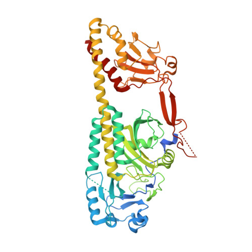
4S21

Crystal structure of the photosensory core module of bacteriophytochrome RPA3015 from R. palustris


Experimental Data Snapshot

  • Method: X-RAY DIFFRACTION
  • Resolution: 3.25 Å
  • R-Value Free: 0.316 
  • R-Value Work: 0.262 
  • R-Value Observed: 0.264 

Starting Model: experimental
View more details

wwPDB Validation   3D Report Full Report


Ligand Structure Quality Assessment 


This is version 1.3 of the entry. See complete history


Literature

Light Signaling Mechanism of Two Tandem Bacteriophytochromes.

Yang, X.Stojkovic, E.A.Ozarowski, W.B.Kuk, J.Davydova, E.Moffat, K.

(2015) Structure 23: 1179-1189

  • DOI: https://doi.org/10.1016/j.str.2015.04.022
  • Primary Citation of Related Structures:  
    4R6L, 4R70, 4S21

  • PubMed Abstract: 

    RpBphP2 and RpBphP3, two tandem bacteriophytochromes from the photosynthetic bacterium Rhodopseudomonas palustris, share high sequence identity but exhibit distinct photoconversion behavior. Unlike the canonical RpBphP2, RpBphP3 photoconverts to an unusual near-red-absorbing (Pnr) state; both are required for synthesis of light-harvesting complexes under low-light conditions. Here we report the crystal structures of the photosensory core modules of RpBphP2 and RpBphP3. Despite different quaternary structures, RpBphP2 and RpBphP3 adopt nearly identical tertiary structures. The RpBphP3 structure reveals tongue-and-groove interactions at the interface between the GAF and PHY domains. A single mutation in the PRxSF motif at the GAF-PHY interface abolishes light-induced formation of the Pnr state in RpBphP3, possibly due to altered structural rigidity of the chromophore-binding pocket. Structural comparisons suggest that long-range signaling involves structural rearrangement of the helical spine at the dimer interface. These structures, together with mutational studies, provide insights into photoconversion and the long-range signaling mechanism in phytochromes.


  • Organizational Affiliation

    Department of Biochemistry and Molecular Biology, University of Chicago, Chicago, IL 60637, USA; Department of Chemistry, University of Illinois at Chicago, Chicago, IL 60607, USA; Department of Ophthalmology and Vision Sciences, University of Illinois at Chicago, Chicago, IL 60607, USA. Electronic address: [email protected].


Macromolecules
Find similar proteins by:  (by identity cutoff)  |  3D Structure
Entity ID: 1
MoleculeChains Sequence LengthOrganismDetailsImage
Bacteriophytochrome (Light-regulated signal transduction histidine kinase), PhyB1
A, B
525Rhodopseudomonas palustris CGA009Mutation(s): 0 
Gene Names: phyB1RPA3015
EC: 2.7.13.3
UniProt
Find proteins for Q6N5G3 (Rhodopseudomonas palustris (strain ATCC BAA-98 / CGA009))
Explore Q6N5G3 
Go to UniProtKB:  Q6N5G3
Entity Groups  
Sequence Clusters30% Identity50% Identity70% Identity90% Identity95% Identity100% Identity
UniProt GroupQ6N5G3
Sequence Annotations
Expand
  • Reference Sequence
Experimental Data & Validation

Experimental Data

  • Method: X-RAY DIFFRACTION
  • Resolution: 3.25 Å
  • R-Value Free: 0.316 
  • R-Value Work: 0.262 
  • R-Value Observed: 0.264 
  • Space Group: P 61
Unit Cell:
Length ( Å )Angle ( ˚ )
a = 175.36α = 90
b = 175.36β = 90
c = 96.19γ = 120
Software Package:
Software NamePurpose
HKL-2000data collection
PHASESphasing
PHENIXrefinement
HKL-2000data reduction
HKL-2000data scaling

Structure Validation

View Full Validation Report


Ligand Structure Quality Assessment 


Entry History 

Deposition Data

Revision History  (Full details and data files)

  • Version 1.0: 2015-07-15
    Type: Initial release
  • Version 1.1: 2015-08-05
    Changes: Database references
  • Version 1.2: 2023-09-20
    Changes: Data collection, Database references, Derived calculations, Refinement description
  • Version 1.3: 2024-10-30
    Changes: Structure summary