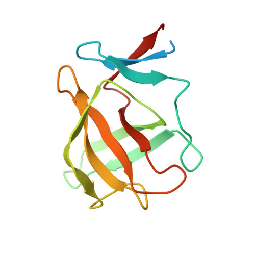
4V30

Cereblon isoform 4 from Magnetospirillum gryphiswaldense in complex with Lenalidomide


Experimental Data Snapshot

  • Method: X-RAY DIFFRACTION
  • Resolution: 1.85 Å
  • R-Value Free: 0.239 
  • R-Value Work: 0.185 
  • R-Value Observed: 0.188 

wwPDB Validation   3D Report Full Report


Ligand Structure Quality Assessment 


This is version 1.1 of the entry. See complete history


Literature

Thalidomide Mimics Uridine Binding to an Aromatic Cage in Cereblon.

Hartmann, M.D.Boichenko, I.Coles, M.Zanini, F.Lupas, A.N.Hernandez Alvarez, B.

(2014) J Struct Biol 188: 225

  • DOI: https://doi.org/10.1016/j.jsb.2014.10.010
  • Primary Citation of Related Structures:  
    4V2Y, 4V2Z, 4V30, 4V31, 4V32

  • PubMed Abstract: 

    Thalidomide and its derivatives lenalidomide and pomalidomide are important anticancer agents but can cause severe birth defects via an interaction with the protein cereblon. The ligand-binding domain of cereblon is found, with a high degree of conservation, in both bacteria and eukaryotes. Using a bacterial model system, we reveal the structural determinants of cereblon substrate recognition, based on a series of high-resolution crystal structures. For the first time, we identify a cellular ligand that is universally present: we show that thalidomide and its derivatives mimic and compete for the binding of uridine, and validate these findings in vivo. The nature of the binding pocket, an aromatic cage of three tryptophan residues, further suggests a role in the recognition of cationic ligands. Our results allow for general evaluation of pharmaceuticals for potential cereblon-dependent teratogenicity.


  • Organizational Affiliation

    Department of Protein Evolution, Max Planck Institute for Developmental Biology, 72076 Tübingen, Germany.


Macromolecules
Find similar proteins by:  (by identity cutoff)  |  3D Structure
Entity ID: 1
MoleculeChains Sequence LengthOrganismDetailsImage
CEREBLON ISOFORM 4
A, B, C
125Magnetospirillum gryphiswaldense MSR-1Mutation(s): 0 
UniProt
Find proteins for A4TVL0 (Magnetospirillum gryphiswaldense)
Explore A4TVL0 
Go to UniProtKB:  A4TVL0
Entity Groups  
Sequence Clusters30% Identity50% Identity70% Identity90% Identity95% Identity100% Identity
UniProt GroupA4TVL0
Sequence Annotations
Expand
  • Reference Sequence
Experimental Data & Validation

Experimental Data

  • Method: X-RAY DIFFRACTION
  • Resolution: 1.85 Å
  • R-Value Free: 0.239 
  • R-Value Work: 0.185 
  • R-Value Observed: 0.188 
  • Space Group: P 21 21 21
Unit Cell:
Length ( Å )Angle ( ˚ )
a = 56.682α = 90
b = 59.743β = 90
c = 88.064γ = 90
Software Package:
Software NamePurpose
REFMACrefinement
XDSdata reduction
XDSdata scaling
SHELXphasing

Structure Validation

View Full Validation Report


Ligand Structure Quality Assessment 


Entry History 

Deposition Data

Revision History  (Full details and data files)

  • Version 1.0: 2014-12-17
    Type: Initial release
  • Version 1.1: 2024-05-08
    Changes: Data collection, Database references, Derived calculations, Other