4V4F

The structure of the trp RNA-binding attenuation protein (TRAP) bound to a RNA molecule containing UAGAU repeats


Experimental Data Snapshot

  • Method: X-RAY DIFFRACTION
  • Resolution: 1.90 Å
  • R-Value Free: 0.236 
  • R-Value Work: 0.186 
  • R-Value Observed: 0.186 

Starting Model: experimental
View more details

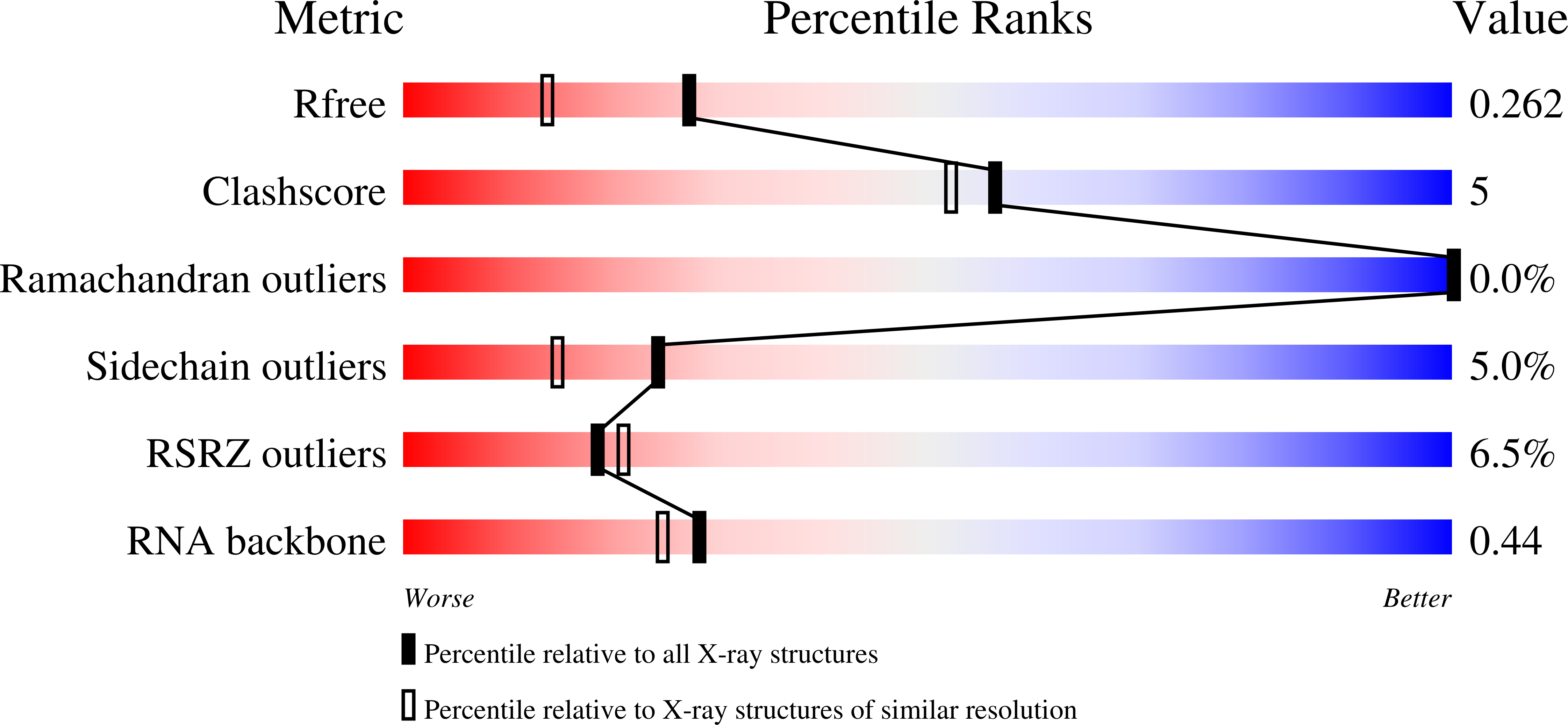
wwPDB Validation   3D Report Full Report


Ligand Structure Quality Assessment 


This is version 1.3 of the entry. See complete history


Literature

The Interaction of RNA with Trap: The Role of Triplet Repeats and Separating Spacer Nucleotides

Hopcroft, N.H.Manfredo, A.Wendt, A.L.Brzozowski, A.M.Gollnick, P.Antson, A.A.

(2004) J Mol Biol 338: 43

  • DOI: https://doi.org/10.1016/j.jmb.2004.02.038
  • Primary Citation of Related Structures:  
    1UTD, 4V4F

  • PubMed Abstract: 

    The trp RNA-binding attenuation protein (TRAP) regulates expression of the tryptophan biosynthetic genes in several Bacilli by binding to the leader region of the nascent trp mRNA, inhibiting continued transcription. The 11 subunit TRAP molecule is active in complex with tryptophan, and binds consequently an RNA target segment consisting of 11 (G/U)AG triplets, each separated by two or three non-conserved "spacer" nucleotides. Here, we report the first crystal structures of TRAP in a complex with RNA containing UAG triplets separated by two nucleotides and in a complex with RNA containing GAG triplets separated by three nucleotides. Comparison with known structures of TRAP-RNA complexes shows that both substitution of G-1 with U-1 in the triplet and addition of an extra spacer nucleotide lead to a more flexible complex. This suggests an explanation why, in the trp leader RNA, all three-nucleotide spacer regions are followed by a G-1 nucleotide. Taken together, the structures demonstrate that RNA binding to TRAP is mediated by specific interactions involving the A-2 and G-3 nucleotides of the triplet. This is accompanied by the disruption of stacking interactions between the bases of the other nucleotides, contributing to the increase in entropy that drives binding.


  • Organizational Affiliation

    York Structural Biology Laboratory, Department of Chemistry, University of York, Heslington, York YO10 5YW, UK.


Macromolecules

Find similar proteins by:  (by identity cutoff)  |  3D Structure
Entity ID: 2
MoleculeChains Sequence LengthOrganismDetailsImage
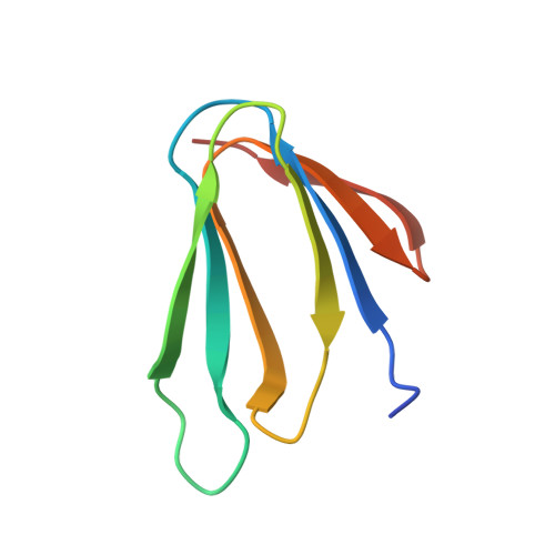
TRANSCRIPTION ATTENUATION PROTEIN MTRB74Geobacillus stearothermophilusMutation(s): 0 
UniProt
Find proteins for Q9X6J6 (Geobacillus stearothermophilus)
Explore Q9X6J6 
Go to UniProtKB:  Q9X6J6
Entity Groups  
Sequence Clusters30% Identity50% Identity70% Identity90% Identity95% Identity100% Identity
UniProt GroupQ9X6J6
Sequence Annotations
Expand
  • Reference Sequence

Find similar nucleic acids by:  Sequence   |   3D Structure  

Entity ID: 1
MoleculeChains LengthOrganismImage
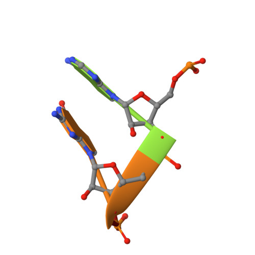
5'-R(*UP*AP*GP*AP*UP)-3'5synthetic construct
Sequence Annotations
Expand
  • Reference Sequence
Small Molecules
Ligands 1 Unique
IDChains Name / Formula / InChI Key2D Diagram3D Interactions
TRP
Query on TRP

Download Ideal Coordinates CCD File 
AC [auth AM]
AD [auth BQ]
BC [auth AN]
BD [auth BR]
CC [auth AO]
AC [auth AM],
AD [auth BQ],
BC [auth AN],
BD [auth BR],
CC [auth AO],
CD [auth BS],
DC [auth AP],
DD [auth BT],
EC [auth AQ],
ED [auth BU],
FC [auth AR],
FD [auth BV],
GC [auth AS],
HC [auth AT],
IC [auth AU],
JC [auth AV],
KC [auth BA],
LC [auth BB],
MC [auth BC],
NC [auth BD],
OB [auth AA],
OC [auth BE],
PB [auth AB],
PC [auth BF],
QB [auth AC],
QC [auth BG],
RB [auth AD],
RC [auth BH],
SB [auth AE],
SC [auth BI],
TB [auth AF],
TC [auth BJ],
UB [auth AG],
UC [auth BK],
VB [auth AH],
VC [auth BL],
WB [auth AI],
WC [auth BM],
XB [auth AJ],
XC [auth BN],
YB [auth AK],
YC [auth BO],
ZB [auth AL],
ZC [auth BP]
TRYPTOPHAN
C11 H12 N2 O2
QIVBCDIJIAJPQS-VIFPVBQESA-N
Experimental Data & Validation

Experimental Data

  • Method: X-RAY DIFFRACTION
  • Resolution: 1.90 Å
  • R-Value Free: 0.236 
  • R-Value Work: 0.186 
  • R-Value Observed: 0.186 
  • Space Group: C 1 2 1
Unit Cell:
Length ( Å )Angle ( ˚ )
a = 113.907α = 90
b = 133.986β = 100.11
c = 232.826γ = 90
Software Package:
Software NamePurpose
AMoREmodel building
REFMACrefinement
DENZOdata reduction
SCALEPACKdata scaling
AMoREphasing

Structure Validation

View Full Validation Report


Ligand Structure Quality Assessment 


Entry History 

Deposition Data

Revision History  (Full details and data files)

  • Version 1.0: 2014-07-09
    Type: Initial release
  • Version 1.1: 2014-12-10
    Changes: Other
  • Version 1.2: 2020-07-29
    Changes: Advisory, Source and taxonomy
  • Version 1.3: 2024-01-10
    Changes: Advisory, Data collection, Database references, Refinement description