4X0W

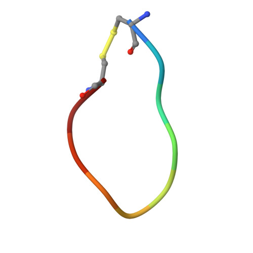
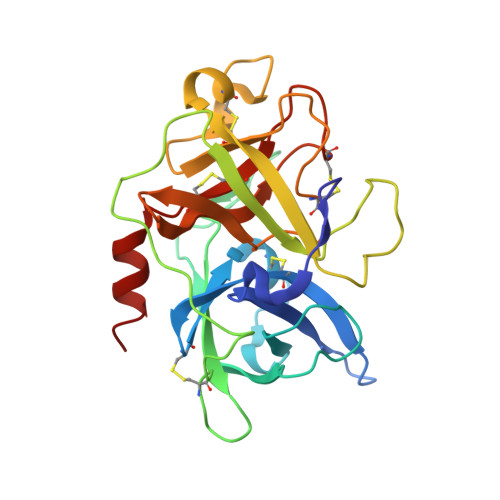
The crystal structure of mupain-1-17 in complex with murinised human uPA


Experimental Data Snapshot

  • Method: X-RAY DIFFRACTION
  • Resolution: 2.10 Å
  • R-Value Free: 0.272 
  • R-Value Work: 0.236 
  • R-Value Observed: 0.238 

wwPDB Validation   3D Report Full Report


This is version 1.1 of the entry. See complete history


Literature

Distinctive binding modes and inhibitory mechanisms of two peptidic inhibitors of urokinase-type plasminogen activator with isomeric P1 residues.

Jiang, L.Zhao, B.Xu, P.Srensen, H.P.Jensen, J.K.Christensen, A.Hosseini, M.Nielsen, N.C.Jensen, K.J.Andreasen, P.A.Huang, M.

(2015) Int J Biochem Cell Biol 62: 88-92

  • DOI: https://doi.org/10.1016/j.biocel.2015.02.016
  • Primary Citation of Related Structures:  
    4X0W, 4X1P

  • PubMed Abstract: 

    Two isomeric piperidine derivatives (meta and para isomers) were used as arginine mimics in the P1 position of a cyclic peptidic inhibitor (CPAYSRYLDC) of urokinase-type plasminogen activator. The two resulting cyclic peptides showed vastly different affinities (∼70 fold) to the target enzyme. X-ray crystal structure analysis showed that the two P1 residues were inserted into the S1 specificity pocket in indistinguishable manners. However, the rest of the peptides bound in entirely different ways on the surface of the enzyme, and the two peptides have different conformations, despite the highly similar sequence. These results demonstrate how the subtle difference in P1 residue can dictate the exosite interactions and the potencies of peptidic inhibitors, and highlight the importance of the P1 residue for protease inhibition. This study provides important information for the development of peptidic agents for pharmacological intervention.


  • Organizational Affiliation

    Fujian Institute of Research on the Structure of Matter, Chinese Academy of Sciences, Fuzhou 350002, China(1); Danish-Chinese Centre for Proteases and Cancer, China.


Macromolecules

Find similar proteins by:  Sequence   |   3D Structure  

Entity ID: 1
MoleculeChains Sequence LengthOrganismDetailsImage
mupain-1-17A [auth P]10synthetic constructMutation(s): 0 
Entity Groups  
Sequence Clusters30% Identity50% Identity70% Identity90% Identity95% Identity100% Identity
Sequence Annotations
Expand
  • Reference Sequence
Find similar proteins by:  (by identity cutoff)  |  3D Structure
Entity ID: 2
MoleculeChains Sequence LengthOrganismDetailsImage
Urokinase-type plasminogen activatorB [auth U]247Homo sapiensMutation(s): 3 
Gene Names: PLAU
EC: 3.4.21.73
UniProt & NIH Common Fund Data Resources
Find proteins for P00749 (Homo sapiens)
Explore P00749 
Go to UniProtKB:  P00749
PHAROS:  P00749
GTEx:  ENSG00000122861 
Entity Groups  
Sequence Clusters30% Identity50% Identity70% Identity90% Identity95% Identity100% Identity
UniProt GroupP00749
Sequence Annotations
Expand
  • Reference Sequence
Experimental Data & Validation

Experimental Data

  • Method: X-RAY DIFFRACTION
  • Resolution: 2.10 Å
  • R-Value Free: 0.272 
  • R-Value Work: 0.236 
  • R-Value Observed: 0.238 
  • Space Group: H 3
Unit Cell:
Length ( Å )Angle ( ˚ )
a = 120.641α = 90
b = 120.641β = 90
c = 42.657γ = 120
Software Package:
Software NamePurpose
REFMACrefinement
Cootmodel building

Structure Validation

View Full Validation Report


Entry History & Funding Information

Deposition Data


Funding OrganizationLocationGrant Number
National Natural Science Foundation of ChinaChina31161130356
National Natural Science Foundation of ChinaChina31170707
Fujian ProvinceChina2012J05071
the Danish National Research FoundationDenmark26-331-6

Revision History  (Full details and data files)

  • Version 1.0: 2015-10-21
    Type: Initial release
  • Version 1.1: 2017-10-18
    Changes: Author supporting evidence, Derived calculations