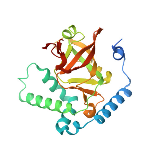
4XZJ

Crystal structure of ADP-ribosyltransferase Vis in complex with NAD


Experimental Data Snapshot

  • Method: X-RAY DIFFRACTION
  • Resolution: 1.80 Å
  • R-Value Free: 0.205 
  • R-Value Work: 0.175 
  • R-Value Observed: 0.177 

Starting Model: experimental
View more details

wwPDB Validation   3D Report Full Report


Ligand Structure Quality Assessment 


This is version 1.4 of the entry. See complete history


Literature

Characterization of Vis Toxin, a Novel ADP-Ribosyltransferase from Vibrio splendidus.

Ravulapalli, R.Lugo, M.R.Pfoh, R.Visschedyk, D.Poole, A.Fieldhouse, R.J.Pai, E.F.Merrill, A.R.

(2015) Biochemistry 54: 5920-5936

  • DOI: https://doi.org/10.1021/acs.biochem.5b00921
  • Primary Citation of Related Structures:  
    4XZJ, 4Y1W, 4YC0

  • PubMed Abstract: 

    Vis toxin was identified by a bioinformatics strategy as a putative virulence factor produced by Vibrio splendidus with mono-ADP-ribosyltransferase activity. Vis was purified to homogeneity as a 28 kDa single-domain enzyme and was shown to possess NAD(+)-glycohydrolase [KM(NAD(+)) = 276 ± 12 μM] activity and with an R-S-E-X-E motif; it targets arginine-related compounds [KM(agmatine) = 272 ± 18 mM]. Mass spectrometry analysis revealed that Vis labels l-arginine with ADP-ribose from the NAD(+) substrate at the amino nitrogen of the guanidinium side chain. Vis is toxic to yeast when expressed in the cytoplasm under control of the CUP1 promotor, and catalytic variants lost the ability to kill the yeast host, indicating that the toxin exerts its lethality through its enzyme activity. Several small molecule inhibitors were identified from a virtual screen, and the most potent compounds were found to inhibit the transferase activity of the enzyme with Ki values ranging from 25 to 134 μM. Inhibitor compound M6 bears the necessary attributes of a solid candidate as a lead compound for therapeutic development. Vis toxin was crystallized, and the structures of the apoenzyme (1.4 Å) and the enzyme bound with NAD(+) (1.8 Å) and with the M6 inhibitor (1.5 Å) were determined. The structures revealed that Vis represents a new subgroup within the mono-ADP-ribosyltransferase toxin family.


  • Organizational Affiliation

    Department of Molecular and Cellular Biology, University of Guelph , Guelph, Ontario, Canada N1G 2W1.


Macromolecules
Find similar proteins by:  (by identity cutoff)  |  3D Structure
Entity ID: 1
MoleculeChains Sequence LengthOrganismDetailsImage
Putative NAD(+)--arginine ADP-ribosyltransferase Vis239Vibrio splendidusMutation(s): 0 
Gene Names: V12B01_18061
EC: 2.4.2.31
UniProt
Find proteins for A3UNN4 (Vibrio splendidus (strain 12B01))
Explore A3UNN4 
Go to UniProtKB:  A3UNN4
Entity Groups  
Sequence Clusters30% Identity50% Identity70% Identity90% Identity95% Identity100% Identity
UniProt GroupA3UNN4
Sequence Annotations
Expand
  • Reference Sequence
Small Molecules
Ligands 1 Unique
IDChains Name / Formula / InChI Key2D Diagram3D Interactions
NAD
Query on NAD

Download Ideal Coordinates CCD File 
B [auth A]NICOTINAMIDE-ADENINE-DINUCLEOTIDE
C21 H27 N7 O14 P2
BAWFJGJZGIEFAR-NNYOXOHSSA-N
Experimental Data & Validation

Experimental Data

  • Method: X-RAY DIFFRACTION
  • Resolution: 1.80 Å
  • R-Value Free: 0.205 
  • R-Value Work: 0.175 
  • R-Value Observed: 0.177 
  • Space Group: P 21 21 21
Unit Cell:
Length ( Å )Angle ( ˚ )
a = 49.7α = 90
b = 52β = 90
c = 100.4γ = 90
Software Package:
Software NamePurpose
XDSdata reduction
PDB_EXTRACTdata extraction
REFMACrefinement
SHELXphasing
PHASERphasing
XPREPdata reduction

Structure Validation

View Full Validation Report


Ligand Structure Quality Assessment 


Entry History 

Deposition Data

Revision History  (Full details and data files)

  • Version 1.0: 2015-09-23
    Type: Initial release
  • Version 1.1: 2015-09-30
    Changes: Database references
  • Version 1.2: 2015-10-28
    Changes: Database references
  • Version 1.3: 2017-11-22
    Changes: Database references, Derived calculations, Refinement description
  • Version 1.4: 2023-09-27
    Changes: Data collection, Database references, Refinement description