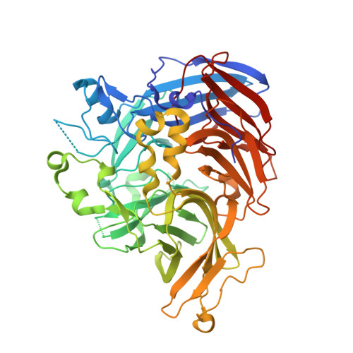
4ZHK

Crystal structure of RPE65 in complex with MB-002

  • Classification: Hydrolase,Isomerase
  • Organism(s): Bos taurus
  • Mutation(s): Yes 
  • Membrane Protein: Yes  OPM

  • Deposited: 2015-04-25 Released: 2015-05-27 
  • Deposition Author(s): Kiser, P.D., Palczewski, K.
  • Funding Organization(s): National Institutes of Health/National Eye Institute (NIH/NEI), Department of Veterans Affairs

Experimental Data Snapshot

  • Method: X-RAY DIFFRACTION
  • Resolution: 2.09 Å
  • R-Value Free: 0.205 
  • R-Value Work: 0.172 
  • R-Value Observed: 0.174 

Starting Model: experimental
View more details

wwPDB Validation   3D Report Full Report


Ligand Structure Quality Assessment 


This is version 1.5 of the entry. See complete history


Literature

Molecular pharmacodynamics of emixustat in protection against retinal degeneration.

Zhang, J.Kiser, P.D.Badiee, M.Palczewska, G.Dong, Z.Golczak, M.Tochtrop, G.P.Palczewski, K.

(2015) J Clin Invest 125: 2781-2794

  • DOI: https://doi.org/10.1172/JCI80950
  • Primary Citation of Related Structures:  
    4RYX, 4RYY, 4RYZ, 4ZHK

  • PubMed Abstract: 

    Emixustat is a visual cycle modulator that has entered clinical trials as a treatment for age-related macular degeneration (AMD). This molecule has been proposed to inhibit the visual cycle isomerase RPE65, thereby slowing regeneration of 11-cis-retinal and reducing production of retinaldehyde condensation byproducts that may be involved in AMD pathology. Previously, we reported that all-trans-retinal (atRAL) is directly cytotoxic and that certain primary amine compounds that transiently sequester atRAL via Schiff base formation ameliorate retinal degeneration. Here, we have shown that emixustat stereoselectively inhibits RPE65 by direct active site binding. However, we detected the presence of emixustat-atRAL Schiff base conjugates, indicating that emixustat also acts as a retinal scavenger, which may contribute to its therapeutic effects. Using agents that lack either RPE65 inhibitory activity or the capacity to sequester atRAL, we assessed the relative importance of these 2 modes of action in protection against retinal phototoxicity in mice. The atRAL sequestrant QEA-B-001-NH2 conferred protection against phototoxicity without inhibiting RPE65, whereas an emixustat derivative incapable of atRAL sequestration was minimally protective, despite direct inhibition of RPE65. These data indicate that atRAL sequestration is an essential mechanism underlying the protective effects of emixustat and related compounds against retinal phototoxicity. Moreover, atRAL sequestration should be considered in the design of next-generation visual cycle modulators.


Macromolecules
Find similar proteins by:  (by identity cutoff)  |  3D Structure
Entity ID: 1
MoleculeChains Sequence LengthOrganismDetailsImage
Retinoid isomerohydrolase
A, B
533Bos taurusMutation(s): 1 
EC: 3.1.1.64 (PDB Primary Data), 5.3.3.22 (UniProt)
Membrane Entity: Yes 
UniProt
Find proteins for Q28175 (Bos taurus)
Explore Q28175 
Go to UniProtKB:  Q28175
Entity Groups  
Sequence Clusters30% Identity50% Identity70% Identity90% Identity95% Identity100% Identity
UniProt GroupQ28175
Sequence Annotations
Expand
  • Reference Sequence
Small Molecules
Ligands 6 Unique
IDChains Name / Formula / InChI Key2D Diagram3D Interactions
A8V
Query on A8V

Download Ideal Coordinates CCD File 
H [auth A],
N [auth B]
(1S)-1-[3-(cyclohexylmethoxy)phenyl]propane-1,3-diol
C16 H24 O3
JEPZJYPEDPLQFU-INIZCTEOSA-N
A9V
Query on A9V

Download Ideal Coordinates CCD File 
I [auth A],
O [auth B]
(1R)-1-[3-(cyclohexylmethoxy)phenyl]propane-1,3-diol
C16 H24 O3
JEPZJYPEDPLQFU-MRXNPFEDSA-N
PG4
Query on PG4

Download Ideal Coordinates CCD File 
D [auth A],
K [auth B]
TETRAETHYLENE GLYCOL
C8 H18 O5
UWHCKJMYHZGTIT-UHFFFAOYSA-N
PGE
Query on PGE

Download Ideal Coordinates CCD File 
F [auth A],
G [auth A],
M [auth B]
TRIETHYLENE GLYCOL
C6 H14 O4
ZIBGPFATKBEMQZ-UHFFFAOYSA-N
FE2
Query on FE2

Download Ideal Coordinates CCD File 
C [auth A],
J [auth B]
FE (II) ION
Fe
CWYNVVGOOAEACU-UHFFFAOYSA-N
NA
Query on NA

Download Ideal Coordinates CCD File 
E [auth A],
L [auth B]
SODIUM ION
Na
FKNQFGJONOIPTF-UHFFFAOYSA-N
Experimental Data & Validation

Experimental Data

  • Method: X-RAY DIFFRACTION
  • Resolution: 2.09 Å
  • R-Value Free: 0.205 
  • R-Value Work: 0.172 
  • R-Value Observed: 0.174 
  • Space Group: P 65
Unit Cell:
Length ( Å )Angle ( ˚ )
a = 175.668α = 90
b = 175.668β = 90
c = 86.297γ = 120
Software Package:
Software NamePurpose
REFMACrefinement
XDSdata reduction
XDSdata scaling
REFMACphasing

Structure Validation

View Full Validation Report


Ligand Structure Quality Assessment 


Entry History & Funding Information

Deposition Data


Funding OrganizationLocationGrant Number
National Institutes of Health/National Eye Institute (NIH/NEI)United StatesEY009339
Department of Veterans AffairsUnited StatesBX002683

Revision History  (Full details and data files)

  • Version 1.0: 2015-05-27
    Type: Initial release
  • Version 1.1: 2017-09-27
    Changes: Advisory, Author supporting evidence, Derived calculations, Source and taxonomy, Structure summary
  • Version 1.2: 2019-12-11
    Changes: Author supporting evidence
  • Version 1.3: 2021-07-07
    Changes: Database references, Derived calculations
  • Version 1.4: 2023-09-27
    Changes: Data collection, Database references, Refinement description
  • Version 1.5: 2024-11-06
    Changes: Structure summary