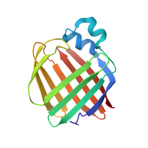
4ZJ0

The crystal structure of monomer Q108K:K40L:Y60W CRBPII bound to all-trans-retinal


Experimental Data Snapshot

  • Method: X-RAY DIFFRACTION
  • Resolution: 1.50 Å
  • R-Value Free: 0.251 
  • R-Value Work: 0.198 
  • R-Value Observed: 0.201 

Starting Model: experimental
View more details

wwPDB Validation   3D Report Full Report


Ligand Structure Quality Assessment 


This is version 1.3 of the entry. See complete history


Literature

Domain-Swapped Dimers of Intracellular Lipid-Binding Proteins: Evidence for Ordered Folding Intermediates.

Assar, Z.Nossoni, Z.Wang, W.Santos, E.M.Kramer, K.McCornack, C.Vasileiou, C.Borhan, B.Geiger, J.H.

(2016) Structure 24: 1590-1598

  • DOI: https://doi.org/10.1016/j.str.2016.05.022
  • Primary Citation of Related Structures:  
    4ZCB, 4ZGU, 4ZH6, 4ZH9, 4ZJ0, 4ZR2, 5DG4, 5DPQ

  • PubMed Abstract: 

    Human Cellular Retinol Binding Protein II (hCRBPII), a member of the intracellular lipid-binding protein family, is a monomeric protein responsible for the intracellular transport of retinol and retinal. Herein we report that hCRBPII forms an extensive domain-swapped dimer during bacterial expression. The domain-swapped region encompasses almost half of the protein. The dimer represents a novel structural architecture with the mouths of the two binding cavities facing each other, producing a new binding cavity that spans the length of the protein complex. Although wild-type hCRBPII forms the dimer, the propensity for dimerization can be substantially increased via mutation at Tyr60. The monomeric form of the wild-type protein represents the thermodynamically more stable species, making the domain-swapped dimer a kinetically trapped entity. Hypothetically, the wild-type protein has evolved to minimize dimerization of the folding intermediate through a critical hydrogen bond (Tyr60-Glu72) that disfavors the dimeric form.


  • Organizational Affiliation

    Department of Chemistry, Michigan State University, East Lansing, MI 48824, USA.


Macromolecules
Find similar proteins by:  (by identity cutoff)  |  3D Structure
Entity ID: 1
MoleculeChains Sequence LengthOrganismDetailsImage
Retinol-binding protein 2
A, B
133Homo sapiensMutation(s): 3 
Gene Names: RBP2CRBP2
UniProt & NIH Common Fund Data Resources
Find proteins for P50120 (Homo sapiens)
Explore P50120 
Go to UniProtKB:  P50120
PHAROS:  P50120
GTEx:  ENSG00000114113 
Entity Groups  
Sequence Clusters30% Identity50% Identity70% Identity90% Identity95% Identity100% Identity
UniProt GroupP50120
Sequence Annotations
Expand
  • Reference Sequence
Experimental Data & Validation

Experimental Data

  • Method: X-RAY DIFFRACTION
  • Resolution: 1.50 Å
  • R-Value Free: 0.251 
  • R-Value Work: 0.198 
  • R-Value Observed: 0.201 
  • Space Group: P 1
Unit Cell:
Length ( Å )Angle ( ˚ )
a = 30.081α = 90.94
b = 36.049β = 91.99
c = 63.92γ = 113.93
Software Package:
Software NamePurpose
REFMACrefinement
DENZOdata reduction
HKL-2000data scaling
REFMACphasing

Structure Validation

View Full Validation Report


Ligand Structure Quality Assessment 


Entry History & Funding Information

Deposition Data


Funding OrganizationLocationGrant Number
National Institutes of Health/National Institute of General Medical Sciences (NIH/NIGMS)United StatesGM101353

Revision History  (Full details and data files)

  • Version 1.0: 2016-05-11
    Type: Initial release
  • Version 1.1: 2017-09-06
    Changes: Author supporting evidence, Database references, Derived calculations
  • Version 1.2: 2019-12-25
    Changes: Author supporting evidence
  • Version 1.3: 2023-09-27
    Changes: Data collection, Database references, Refinement description