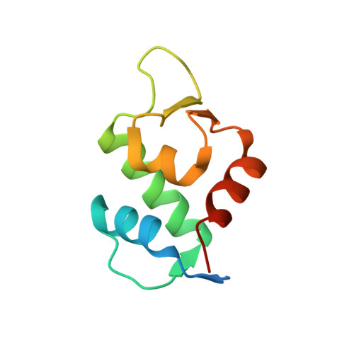
4ZYC

Discovery of dihydroisoquinolinone derivatives as novel inhibitors of the p53-MDM2 interaction with a distinct binding mode: Hdm2 (MDM2) complexed with cpd5


Experimental Data Snapshot

  • Method: X-RAY DIFFRACTION
  • Resolution: 1.95 Å
  • R-Value Free: 0.259 
  • R-Value Work: 0.220 
  • R-Value Observed: 0.222 

Starting Model: experimental
View more details

wwPDB Validation   3D Report Full Report


Ligand Structure Quality Assessment 


This is version 1.3 of the entry. See complete history


Literature

Discovery of dihydroisoquinolinone derivatives as novel inhibitors of the p53-MDM2 interaction with a distinct binding mode.

Gessier, F.Kallen, J.Jacoby, E.Chene, P.Stachyra-Valat, T.Ruetz, S.Jeay, S.Holzer, P.Masuya, K.Furet, P.

(2015) Bioorg Med Chem Lett 25: 3621-3625

  • DOI: https://doi.org/10.1016/j.bmcl.2015.06.058
  • Primary Citation of Related Structures:  
    4ZYC

  • PubMed Abstract: 

    Blocking the interaction between the p53 tumor suppressor and its regulatory protein MDM2 is a promising therapeutic concept under current investigation in oncology drug research. We report here the discovery of the first representatives of a new class of small molecule inhibitors of this protein-protein interaction: the dihydroisoquinolinones. Starting from an initial hit identified by virtual screening, a derivatization program has resulted in compound 11, a low nanomolar inhibitor of the p53-MDM2 interaction showing significant cellular activity. Initially based on a binding mode hypothesis, this effort was then guided by a X-ray co-crystal structure of MDM2 in complex with one of the synthesized analogs. The X-ray structure revealed an unprecedented binding mode for p53-MDM2 inhibitors.


  • Organizational Affiliation

    Novartis Institutes for BioMedical Research, WKL-136.P.12, CH-4002 Basel, Switzerland.


Macromolecules
Find similar proteins by:  (by identity cutoff)  |  3D Structure
Entity ID: 1
MoleculeChains Sequence LengthOrganismDetailsImage
E3 ubiquitin-protein ligase Mdm2
A, B, C
96Homo sapiensMutation(s): 1 
Gene Names: MDM2
EC: 6.3.2 (PDB Primary Data), 2.3.2.27 (UniProt)
UniProt & NIH Common Fund Data Resources
Find proteins for Q00987 (Homo sapiens)
Explore Q00987 
Go to UniProtKB:  Q00987
PHAROS:  Q00987
GTEx:  ENSG00000135679 
Entity Groups  
Sequence Clusters30% Identity50% Identity70% Identity90% Identity95% Identity100% Identity
UniProt GroupQ00987
Sequence Annotations
Expand
  • Reference Sequence
Experimental Data & Validation

Experimental Data

  • Method: X-RAY DIFFRACTION
  • Resolution: 1.95 Å
  • R-Value Free: 0.259 
  • R-Value Work: 0.220 
  • R-Value Observed: 0.222 
  • Space Group: P 1 21 1
Unit Cell:
Length ( Å )Angle ( ˚ )
a = 39.02α = 90
b = 49.78β = 92.77
c = 75.287γ = 90
Software Package:
Software NamePurpose
DENZOdata reduction
SCALEPACKdata scaling
MOLREPphasing
REFMACrefinement
PDB_EXTRACTdata extraction

Structure Validation

View Full Validation Report


Ligand Structure Quality Assessment 


Entry History 

Deposition Data

  • Released Date: 2015-07-22 
  • Deposition Author(s): Kallen, J.

Revision History  (Full details and data files)

  • Version 1.0: 2015-07-22
    Type: Initial release
  • Version 1.1: 2015-08-12
    Changes: Database references
  • Version 1.2: 2017-11-29
    Changes: Database references
  • Version 1.3: 2024-01-10
    Changes: Data collection, Database references, Refinement description