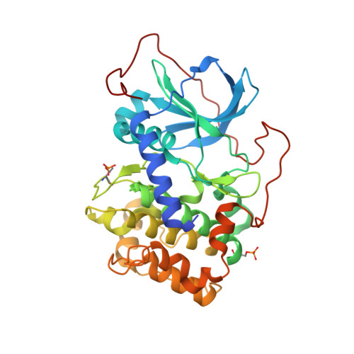
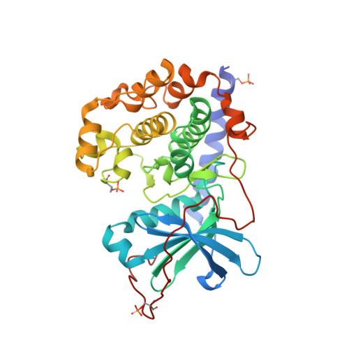
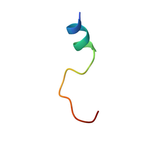
4WB6

Crystal structure of a L205R mutant of human cAMP-dependent protein kinase A (catalytic alpha subunit)


Experimental Data Snapshot

  • Method: X-RAY DIFFRACTION
  • Resolution: 2.10 Å
  • R-Value Free: 0.237 
  • R-Value Work: 0.188 
  • R-Value Observed: 0.190 

Starting Model: experimental
View more details

wwPDB Validation   3D Report Full Report


Ligand Structure Quality Assessment 


This is version 1.4 of the entry. See complete history


Literature

Structural insights into mis-regulation of protein kinase A in human tumors.

Cheung, J.Ginter, C.Cassidy, M.Franklin, M.C.Rudolph, M.J.Robine, N.Darnell, R.B.Hendrickson, W.A.

(2015) Proc Natl Acad Sci U S A 112: 1374-1379

  • DOI: https://doi.org/10.1073/pnas.1424206112
  • Primary Citation of Related Structures:  
    4WB5, 4WB6, 4WB7, 4WB8

  • PubMed Abstract: 

    The extensively studied cAMP-dependent protein kinase A (PKA) is involved in the regulation of critical cell processes, including metabolism, gene expression, and cell proliferation; consequentially, mis-regulation of PKA signaling is implicated in tumorigenesis. Recent genomic studies have identified recurrent mutations in the catalytic subunit of PKA in tumors associated with Cushing's syndrome, a kidney disorder leading to excessive cortisol production, and also in tumors associated with fibrolamellar hepatocellular carcinoma (FL-HCC), a rare liver cancer. Expression of a L205R point mutant and a DnaJ-PKA fusion protein were found to be linked to Cushing's syndrome and FL-HCC, respectively. Here we reveal contrasting mechanisms for increased PKA signaling at the molecular level through structural determination and biochemical characterization of the aberrant enzymes. In the Cushing's syndrome disorder, we find that the L205R mutation abolishes regulatory-subunit binding, leading to constitutive, cAMP-independent signaling. In FL-HCC, the DnaJ-PKA chimera remains under regulatory subunit control; however, its overexpression from the DnaJ promoter leads to enhanced cAMP-dependent signaling. Our findings provide a structural understanding of the two distinct disease mechanisms and they offer a basis for designing effective drugs for their treatment.


  • Organizational Affiliation

    New York Structural Biology Center, New York, NY 10027; [email protected] [email protected].


Macromolecules
Find similar proteins by:  (by identity cutoff)  |  3D Structure
Entity ID: 1
MoleculeChains Sequence LengthOrganismDetailsImage
cAMP-dependent protein kinase catalytic subunit alpha350Homo sapiensMutation(s): 1 
Gene Names: PRKACAPKACA
EC: 2.7.11.11
UniProt & NIH Common Fund Data Resources
Find proteins for P17612 (Homo sapiens)
Explore P17612 
Go to UniProtKB:  P17612
PHAROS:  P17612
GTEx:  ENSG00000072062 
Entity Groups  
Sequence Clusters30% Identity50% Identity70% Identity90% Identity95% Identity100% Identity
UniProt GroupP17612
Sequence Annotations
Expand
  • Reference Sequence
Find similar proteins by:  (by identity cutoff)  |  3D Structure
Entity ID: 2
MoleculeChains Sequence LengthOrganismDetailsImage
cAMP-dependent protein kinase catalytic subunit alpha350Homo sapiensMutation(s): 1 
Gene Names: PRKACAPKACA
EC: 2.7.11.11
UniProt & NIH Common Fund Data Resources
Find proteins for P17612 (Homo sapiens)
Explore P17612 
Go to UniProtKB:  P17612
PHAROS:  P17612
GTEx:  ENSG00000072062 
Entity Groups  
Sequence Clusters30% Identity50% Identity70% Identity90% Identity95% Identity100% Identity
UniProt GroupP17612
Sequence Annotations
Expand
  • Reference Sequence

Find similar proteins by:  Sequence   |   3D Structure  

Entity ID: 3
MoleculeChains Sequence LengthOrganismDetailsImage
PKI (5-24)C [auth I],
D [auth J]
20Homo sapiensMutation(s): 0 
UniProt & NIH Common Fund Data Resources
Find proteins for P61925 (Homo sapiens)
Explore P61925 
Go to UniProtKB:  P61925
PHAROS:  P61925
GTEx:  ENSG00000171033 
Entity Groups  
Sequence Clusters30% Identity50% Identity70% Identity90% Identity95% Identity100% Identity
UniProt GroupP61925
Sequence Annotations
Expand
  • Reference Sequence
Experimental Data & Validation

Experimental Data

  • Method: X-RAY DIFFRACTION
  • Resolution: 2.10 Å
  • R-Value Free: 0.237 
  • R-Value Work: 0.188 
  • R-Value Observed: 0.190 
  • Space Group: P 1 21 1
Unit Cell:
Length ( Å )Angle ( ˚ )
a = 56.169α = 90
b = 90.193β = 96.12
c = 90.377γ = 90
Software Package:
Software NamePurpose
PHENIXrefinement

Structure Validation

View Full Validation Report


Ligand Structure Quality Assessment 


Entry History 

Deposition Data

Revision History  (Full details and data files)

  • Version 1.0: 2015-01-21
    Type: Initial release
  • Version 1.1: 2015-02-04
    Changes: Database references
  • Version 1.2: 2015-02-11
    Changes: Database references
  • Version 1.3: 2023-09-27
    Changes: Data collection, Database references, Derived calculations, Other, Refinement description, Source and taxonomy, Structure summary
  • Version 1.4: 2024-10-23
    Changes: Structure summary