5A5K

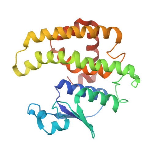
AtGSTF2 from Arabidopsis thaliana in complex with camalexin


Experimental Data Snapshot

  • Method: X-RAY DIFFRACTION
  • Resolution: 2.77 Å
  • R-Value Free: 0.284 
  • R-Value Work: 0.250 
  • R-Value Observed: 0.252 

Starting Model: experimental
View more details

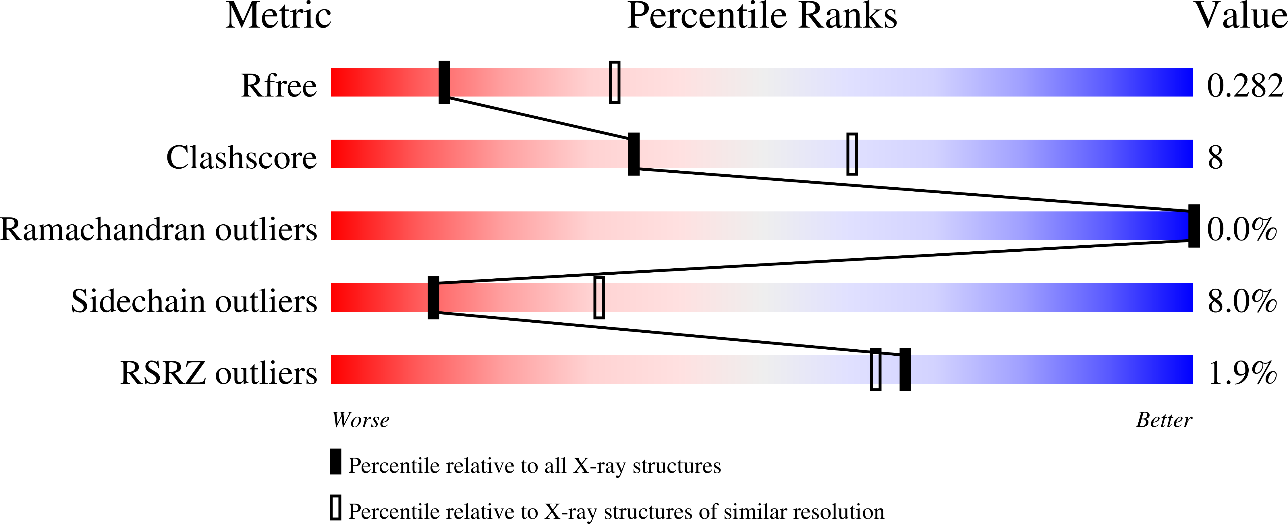
wwPDB Validation   3D Report Full Report


Ligand Structure Quality Assessment 


This is version 1.3 of the entry. See complete history


Literature

Structural evidence for Arabidopsis glutathione transferase AtGSTF2 functioning as a transporter of small organic ligands.

Ahmad, L.Rylott, E.L.Bruce, N.C.Edwards, R.Grogan, G.

(2017) FEBS Open Bio 7: 122-132

  • DOI: https://doi.org/10.1002/2211-5463.12168
  • Primary Citation of Related Structures:  
    5A4U, 5A4V, 5A4W, 5A5K

  • PubMed Abstract: 

    Glutathione transferases (GSTs) are involved in many processes in plant biochemistry, with their best characterised role being the detoxification of xenobiotics through their conjugation with glutathione. GSTs have also been implicated in noncatalytic roles, including the binding and transport of small heterocyclic ligands such as indole hormones, phytoalexins and flavonoids. Although evidence for ligand binding and transport has been obtained using gene deletions and ligand binding studies on purified GSTs, there has been no structural evidence for the binding of relevant ligands in noncatalytic sites. Here we provide evidence of noncatalytic ligand-binding sites in the phi class GST from the model plant Arabidopsis thaliana , At GSTF2, revealed by X-ray crystallography. Complexes of the At GSTF2 dimer were obtained with indole-3-aldehyde, camalexin, the flavonoid quercetrin and its non-rhamnosylated analogue quercetin, at resolutions of 2.00, 2.77, 2.25 and 2.38 Å respectively. Two symmetry-equivalent-binding sites ( L1 ) were identified at the periphery of the dimer, and one more ( L2 ) at the dimer interface. In the complexes, indole-3-aldehyde and quercetrin were found at both L1 and L2 sites, but camalexin was found only at the L1 sites and quercetin only at the L2 site. Ligand binding at each site appeared to be largely determined through hydrophobic interactions. The crystallographic studies support previous conclusions made on ligand binding in noncatalytic sites by At GSTF2 based on isothermal calorimetry experiments (Dixon et al . (2011) Biochem J 438 , 63-70) and suggest a mode of ligand binding in GSTs commensurate with a possible role in ligand transport.


  • Organizational Affiliation

    York Structural Biology Laboratory Department of Chemistry University of York UK; Department of Biology Centre for Novel Agricultural Products University of York UK.


Macromolecules
Find similar proteins by:  (by identity cutoff)  |  3D Structure
Entity ID: 1
MoleculeChains Sequence LengthOrganismDetailsImage
GLUTATHIONE S-TRANSFERASE F2
A, B, C, D, E
A, B, C, D, E, F, G, H, I, J, K, L, M, N, O, P, Q, R, S, T, U, V, W, X
212Arabidopsis thalianaMutation(s): 0 
EC: 2.5.1.18
UniProt
Find proteins for P46422 (Arabidopsis thaliana)
Explore P46422 
Go to UniProtKB:  P46422
Entity Groups  
Sequence Clusters30% Identity50% Identity70% Identity90% Identity95% Identity100% Identity
UniProt GroupP46422
Sequence Annotations
Expand
  • Reference Sequence
Small Molecules
Ligands 1 Unique
IDChains Name / Formula / InChI Key2D Diagram3D Interactions
7WB
Query on 7WB

Download Ideal Coordinates CCD File 
AA [auth C]
BA [auth D]
CA [auth E]
DA [auth F]
EA [auth G]
AA [auth C],
BA [auth D],
CA [auth E],
DA [auth F],
EA [auth G],
FA [auth H],
GA [auth I],
HA [auth J],
IA [auth K],
JA [auth L],
KA [auth M],
LA [auth N],
MA [auth O],
NA [auth P],
OA [auth Q],
PA [auth R],
QA [auth S],
RA [auth T],
SA [auth U],
TA [auth V],
UA [auth W],
VA [auth X],
Y [auth A],
Z [auth B]
(2Z)-2-indol-3-ylidene-3H-1,3-thiazole
C11 H8 N2 S1
IUBODNCSFJZWGR-PKNBQFBNSA-N
Experimental Data & Validation

Experimental Data

  • Method: X-RAY DIFFRACTION
  • Resolution: 2.77 Å
  • R-Value Free: 0.284 
  • R-Value Work: 0.250 
  • R-Value Observed: 0.252 
  • Space Group: P 1
Unit Cell:
Length ( Å )Angle ( ˚ )
a = 97.1α = 83.65
b = 113.72β = 79.47
c = 132.02γ = 65.85
Software Package:
Software NamePurpose
REFMACrefinement
XDSdata reduction
SCALAdata scaling
MOLREPphasing

Structure Validation

View Full Validation Report


Ligand Structure Quality Assessment 


Entry History 

Deposition Data

Revision History  (Full details and data files)

  • Version 1.0: 2016-06-29
    Type: Initial release
  • Version 1.1: 2017-01-11
    Changes: Database references
  • Version 1.2: 2017-03-01
    Changes: Database references
  • Version 1.3: 2024-01-10
    Changes: Data collection, Database references, Derived calculations, Other, Refinement description