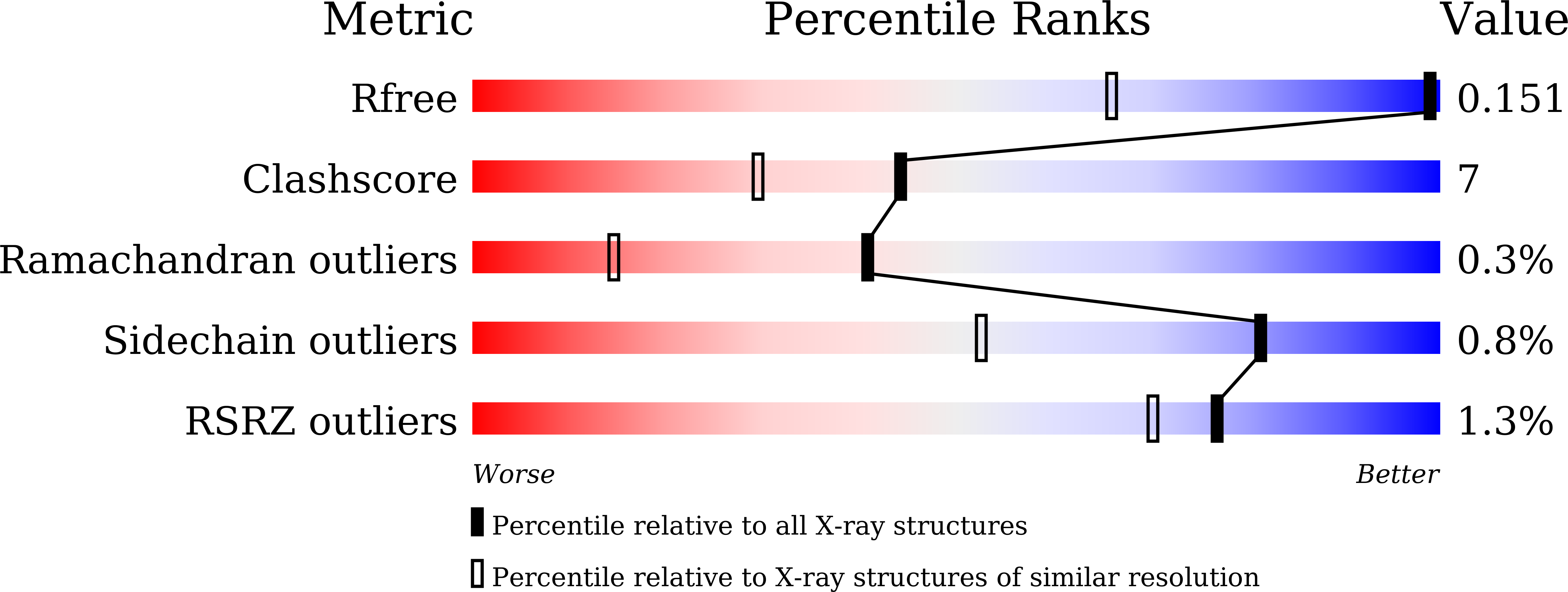
5AVN

The 1.03 angstrom structure (P212121) of glucose isomerase crystallized in high-strength agarose hydrogel


Experimental Data Snapshot

  • Method: X-RAY DIFFRACTION
  • Resolution: 1.03 Å
  • R-Value Free: 0.150 
  • R-Value Work: 0.127 
  • R-Value Observed: 0.129 

Starting Model: experimental
View more details

wwPDB Validation   3D Report Full Report


This is version 1.2 of the entry. See complete history


Literature

Growth of protein crystals in hydrogels prevents osmotic shock

Sugiyama, S.Maruyama, M.Sazaki, G.Hirose, M.Adachi, H.Takano, K.Murakami, S.Inoue, T.Mori, Y.Matsumura, H.

(2012) J Am Chem Soc 134: 5786-5789

  • DOI: https://doi.org/10.1021/ja301584y
  • Primary Citation of Related Structures:  
    5AVD, 5AVG, 5AVH, 5AVN

  • PubMed Abstract: 

    High-throughput protein X-ray crystallography offers a significant opportunity to facilitate drug discovery. The most reliable approach is to determine the three-dimensional structure of the protein-ligand complex by soaking the ligand in apo crystals. However, protein apo crystals produced by conventional crystallization in a solution are fatally damaged by osmotic shock during soaking. To overcome this difficulty, we present a novel technique for growing protein crystals in a high-concentration hydrogel that is completely gellified and exhibits high strength. This technique allowed us essentially to increase the mechanical stability of the crystals, preventing serious damage to the crystals caused by osmotic shock. Thus, this method may accelerate structure-based drug discoveries.


  • Organizational Affiliation

    Graduate School of Engineering, Osaka University, Suita, Osaka 565-0871, Japan. [email protected]


Macromolecules
Find similar proteins by:  (by identity cutoff)  |  3D Structure
Entity ID: 1
MoleculeChains Sequence LengthOrganismDetailsImage
Xylose isomerase
A, B
387Streptomyces rubiginosusMutation(s): 0 
Gene Names: xylA
EC: 5.3.1.5
UniProt
Find proteins for P24300 (Streptomyces rubiginosus)
Explore P24300 
Go to UniProtKB:  P24300
Entity Groups  
Sequence Clusters30% Identity50% Identity70% Identity90% Identity95% Identity100% Identity
UniProt GroupP24300
Sequence Annotations
Expand
  • Reference Sequence
Small Molecules
Ligands 3 Unique
IDChains Name / Formula / InChI Key2D Diagram3D Interactions
SO4
Query on SO4

Download Ideal Coordinates CCD File 
E [auth A],
F [auth A],
I [auth B],
J [auth B]
SULFATE ION
O4 S
QAOWNCQODCNURD-UHFFFAOYSA-L
MN
Query on MN

Download Ideal Coordinates CCD File 
C [auth A],
G [auth B]
MANGANESE (II) ION
Mn
WAEMQWOKJMHJLA-UHFFFAOYSA-N
CA
Query on CA

Download Ideal Coordinates CCD File 
D [auth A],
H [auth B]
CALCIUM ION
Ca
BHPQYMZQTOCNFJ-UHFFFAOYSA-N
Experimental Data & Validation

Experimental Data

  • Method: X-RAY DIFFRACTION
  • Resolution: 1.03 Å
  • R-Value Free: 0.150 
  • R-Value Work: 0.127 
  • R-Value Observed: 0.129 
  • Space Group: P 21 21 2
Unit Cell:
Length ( Å )Angle ( ˚ )
a = 94.082α = 90
b = 98.285β = 90
c = 85.777γ = 90
Software Package:
Software NamePurpose
REFMACrefinement
DENZOdata reduction
HKL-2000data scaling
MOLREPphasing

Structure Validation

View Full Validation Report


Entry History & Funding Information

Deposition Data


Funding OrganizationLocationGrant Number
JSPS KAKENHIJapan23860028
JSPS KAKENHIJapan25650051
JSPS KAKENHIJapan25286051

Revision History  (Full details and data files)

  • Version 1.0: 2015-07-08
    Type: Initial release
  • Version 1.1: 2020-02-26
    Changes: Advisory, Data collection, Derived calculations, Source and taxonomy
  • Version 1.2: 2023-11-08
    Changes: Data collection, Database references, Derived calculations, Refinement description