5B03

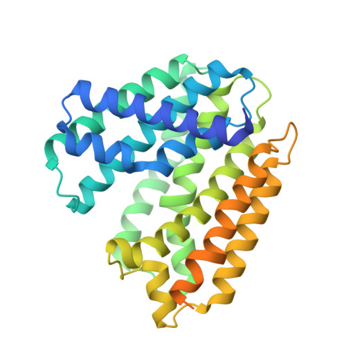
Structure of MoeN5-Sso7d fusion protein in complex with geranyl pyrophosphate


Experimental Data Snapshot

  • Method: X-RAY DIFFRACTION
  • Resolution: 2.60 Å
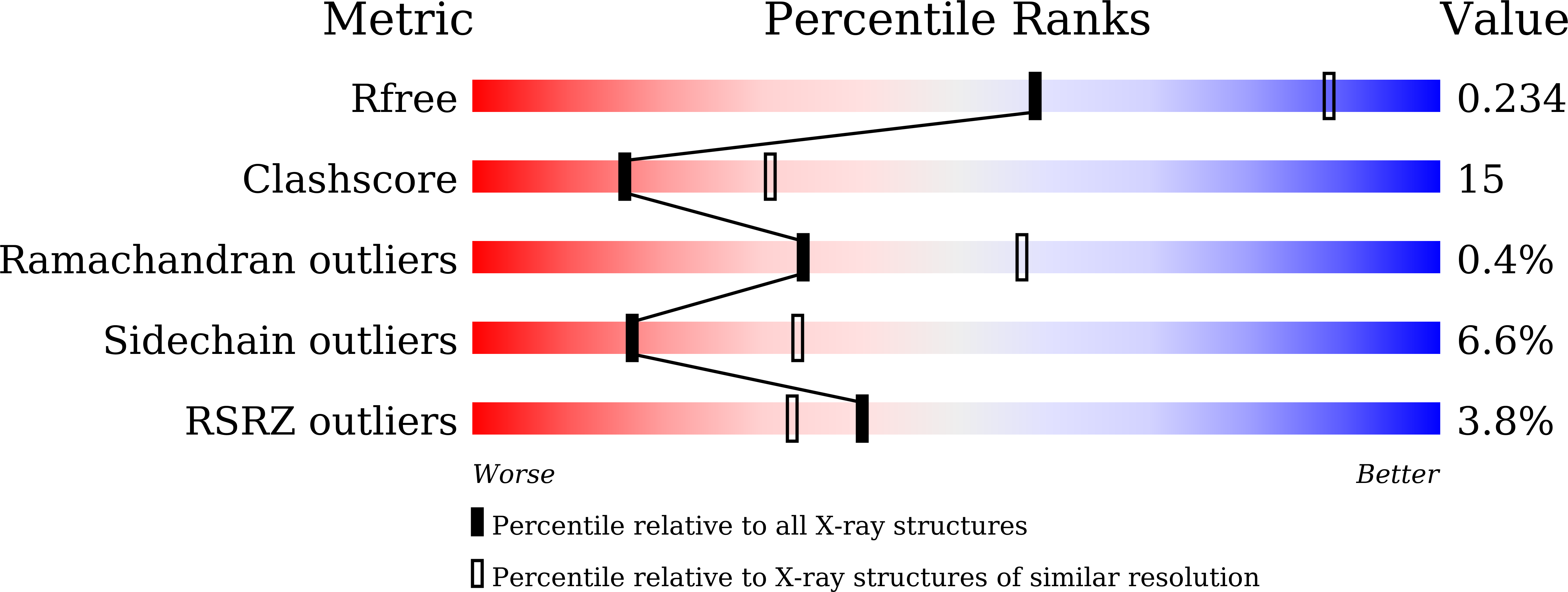
  • R-Value Free: 0.239 
  • R-Value Work: 0.178 

Starting Model: experimental
View more details

wwPDB Validation   3D Report Full Report


Ligand Structure Quality Assessment 


This is version 1.2 of the entry. See complete history


Literature

Moenomycin Biosynthesis: Structure and Mechanism of Action of the Prenyltransferase MoeN5.

Zhang, L.Chen, C.C.Ko, T.P.Huang, J.W.Zheng, Y.Liu, W.Wang, I.Malwal, S.R.Feng, X.Wang, K.Huang, C.H.Hsu, S.T.Wang, A.H.Oldfield, E.Guo, R.T.

(2016) Angew Chem Int Ed Engl 55: 4716-4720

  • DOI: https://doi.org/10.1002/anie.201511388
  • Primary Citation of Related Structures:  
    5B00, 5B02, 5B03, 5B0I, 5B0J, 5B0K, 5B0L, 5B0M

  • PubMed Abstract: 

    The structure of MoeN5, a unique prenyltransferase involved in the biosynthesis of the antibiotic moenomycin, is reported. MoeN5 catalyzes the reaction of geranyl diphosphate (GPP) with the cis-farnesyl group in phosphoglycolipid 5 to form the (C25) moenocinyl-sidechain-containing lipid 7. GPP binds to an allylic site (S1) and aligns well with known S1 inhibitors. Alkyl glycosides, glycolipids, can bind to both S1 and a second site, S2. Long sidechains in S2 are "bent" and co-locate with the homoallylic substrate isopentenyl diphosphate in other prenyltransferases. These observations support a MoeN5 mechanism in which 5 binds to S2 with its C6-C11 group poised to attack C1 in GPP to form the moenocinyl sidechain, with the more distal regions of 5 aligning with the distal glucose in decyl maltoside. The results are of general interest because they provide the first structures of MoeN5 and a structural basis for its mechanism of action, results that will facilitate the design of new antibiotics.


  • Organizational Affiliation

    Industrial Enzymes National Engineering Laboratory, Tianjin Institute of Industrial Biotechnology, Chinese Academy of Sciences, Tianjin, 300308, China.


Macromolecules
Find similar proteins by:  (by identity cutoff)  |  3D Structure
Entity ID: 1
MoleculeChains Sequence LengthOrganismDetailsImage
MoeN5,DNA-binding protein 7d
A, B, C, D
343Streptomyces viridosporusSaccharolobus solfataricus P2
This entity is chimeric
Mutation(s): 0 
Gene Names: moeN5sso7dsso7d-1SSO10610
UniProt
Find proteins for A0A010 (Streptomyces viridosporus)
Explore A0A010 
Go to UniProtKB:  A0A010
Find proteins for P39476 (Saccharolobus solfataricus (strain ATCC 35092 / DSM 1617 / JCM 11322 / P2))
Explore P39476 
Go to UniProtKB:  P39476
Entity Groups  
Sequence Clusters30% Identity50% Identity70% Identity90% Identity95% Identity100% Identity
UniProt GroupsA0A010P39476
Sequence Annotations
Expand
  • Reference Sequence
Experimental Data & Validation

Experimental Data

  • Method: X-RAY DIFFRACTION
  • Resolution: 2.60 Å
  • R-Value Free: 0.239 
  • R-Value Work: 0.178 
  • Space Group: C 2 2 2
Unit Cell:
Length ( Å )Angle ( ˚ )
a = 137.546α = 90
b = 217.336β = 90
c = 104.38γ = 90
Software Package:
Software NamePurpose
CNSrefinement
HKL-2000data reduction
HKL-2000data scaling
CNSphasing
Blu-Icedata collection

Structure Validation

View Full Validation Report


Ligand Structure Quality Assessment 


Entry History 

Deposition Data

Revision History  (Full details and data files)

  • Version 1.0: 2016-03-23
    Type: Initial release
  • Version 1.1: 2016-04-20
    Changes: Database references
  • Version 1.2: 2023-11-08
    Changes: Data collection, Database references, Derived calculations, Refinement description