5CEI

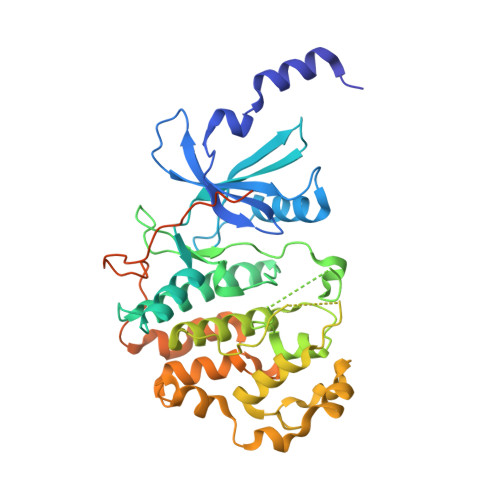
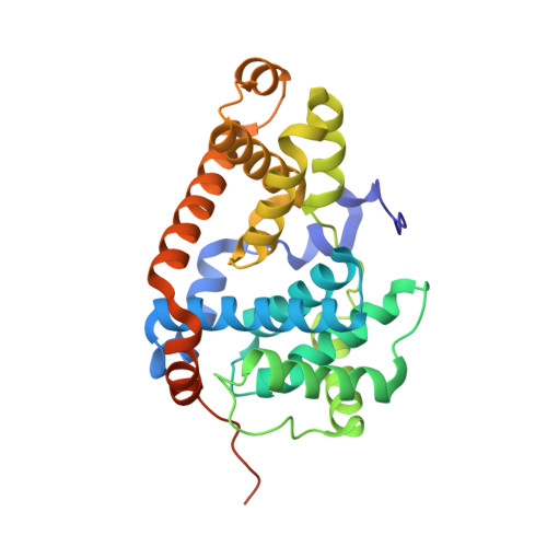
Crystal structure of CDK8:Cyclin C complex with compound 22


Experimental Data Snapshot

  • Method: X-RAY DIFFRACTION
  • Resolution: 2.24 Å
  • R-Value Free: 0.227 
  • R-Value Work: 0.173 
  • R-Value Observed: 0.175 

wwPDB Validation   3D Report Full Report


Ligand Structure Quality Assessment 


This is version 1.2 of the entry. See complete history


Literature

Development of a Potent, Specific CDK8 Kinase Inhibitor Which Phenocopies CDK8/19 Knockout Cells.

Koehler, M.F.Bergeron, P.Blackwood, E.M.Bowman, K.Clark, K.R.Firestein, R.Kiefer, J.R.Maskos, K.McCleland, M.L.Orren, L.Salphati, L.Schmidt, S.Schneider, E.V.Wu, J.Beresini, M.H.

(2016) ACS Med Chem Lett 7: 223-228

  • DOI: https://doi.org/10.1021/acsmedchemlett.5b00278
  • Primary Citation of Related Structures:  
    5CEI

  • PubMed Abstract: 

    Beginning with promiscuous COT inhibitors, which were found to inhibit CDK8, a series of 6-aza-benzothiophene containing compounds were developed into potent, selective CDK8 inhibitors. When cocrystallized with CDK8 and cyclin C, these compounds exhibit an unusual binding mode, making a single hydrogen bond to the hinge residue A100, a second to K252, and a key cation-π interaction with R356. Structure-based drug design resulted in tool compounds 13 and 32, which are highly potent, kinase selective, permeable compounds with a free fraction >2% and no measurable efflux. Despite these attractive properties, these compounds exhibit weak antiproliferative activity in the HCT-116 colon cancer cell line. Further examination of the activity of 32 in this cell line revealed that the compound reduced phosphorylation of the known CDK8 substrate STAT1 in a manner identical to a CDK8 knockout clone, illustrating the complex effects of inhibition of CDK8 kinase activity in proliferation in these cells.


  • Organizational Affiliation

    Departments of Discovery Chemistry, Translational Oncology, Structural Biology, Biochemical and Cellular Pharmacology, Pathology, Drug Metabolism and Pharmacokinetics, and Structural Biology, Genentech, Inc. , 1 DNA Way, South San Francisco, California 94080, United States.


Macromolecules
Find similar proteins by:  (by identity cutoff)  |  3D Structure
Entity ID: 1
MoleculeChains Sequence LengthOrganismDetailsImage
Cyclin-dependent kinase 8406Homo sapiensMutation(s): 0 
Gene Names: CDK8
EC: 2.7.11.22 (PDB Primary Data), 2.7.11.23 (PDB Primary Data)
UniProt & NIH Common Fund Data Resources
Find proteins for P49336 (Homo sapiens)
Explore P49336 
Go to UniProtKB:  P49336
PHAROS:  P49336
GTEx:  ENSG00000132964 
Entity Groups  
Sequence Clusters30% Identity50% Identity70% Identity90% Identity95% Identity100% Identity
UniProt GroupP49336
Sequence Annotations
Expand
  • Reference Sequence
Find similar proteins by:  (by identity cutoff)  |  3D Structure
Entity ID: 2
MoleculeChains Sequence LengthOrganismDetailsImage
Cyclin-C287Homo sapiensMutation(s): 0 
Gene Names: CCNC
UniProt & NIH Common Fund Data Resources
Find proteins for P24863 (Homo sapiens)
Explore P24863 
Go to UniProtKB:  P24863
PHAROS:  P24863
GTEx:  ENSG00000112237 
Entity Groups  
Sequence Clusters30% Identity50% Identity70% Identity90% Identity95% Identity100% Identity
UniProt GroupP24863
Sequence Annotations
Expand
  • Reference Sequence
Small Molecules
Ligands 3 Unique
IDChains Name / Formula / InChI Key2D Diagram3D Interactions
50R
Query on 50R

Download Ideal Coordinates CCD File 
J [auth A]4-(4-iodophenoxy)-N-methylthieno[2,3-c]pyridine-2-carboxamide
C15 H11 I N2 O2 S
QHMOZKVAGIEXJR-UHFFFAOYSA-N
EDO
Query on EDO

Download Ideal Coordinates CCD File 
C [auth A]
D [auth A]
E [auth A]
F [auth A]
K [auth A]
C [auth A],
D [auth A],
E [auth A],
F [auth A],
K [auth A],
L [auth B],
M [auth B],
R [auth B]
1,2-ETHANEDIOL
C2 H6 O2
LYCAIKOWRPUZTN-UHFFFAOYSA-N
FMT
Query on FMT

Download Ideal Coordinates CCD File 
G [auth A]
H [auth A]
I [auth A]
N [auth B]
O [auth B]
G [auth A],
H [auth A],
I [auth A],
N [auth B],
O [auth B],
P [auth B],
Q [auth B]
FORMIC ACID
C H2 O2
BDAGIHXWWSANSR-UHFFFAOYSA-N
Experimental Data & Validation

Experimental Data

  • Method: X-RAY DIFFRACTION
  • Resolution: 2.24 Å
  • R-Value Free: 0.227 
  • R-Value Work: 0.173 
  • R-Value Observed: 0.175 
  • Space Group: P 21 21 21
Unit Cell:
Length ( Å )Angle ( ˚ )
a = 71.128α = 90
b = 70.09β = 90
c = 167.448γ = 90
Software Package:
Software NamePurpose
PHENIXrefinement
SCALAdata scaling
PDB_EXTRACTdata extraction
XDSdata reduction
PHASERphasing

Structure Validation

View Full Validation Report


Ligand Structure Quality Assessment 


Entry History 

Deposition Data

Revision History  (Full details and data files)

  • Version 1.0: 2016-02-10
    Type: Initial release
  • Version 1.1: 2016-05-11
    Changes: Database references
  • Version 1.2: 2024-03-06
    Changes: Data collection, Database references, Derived calculations