5CTG

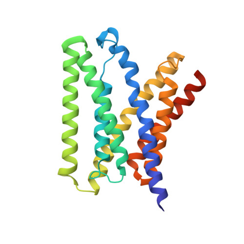
The 3.1 A resolution structure of a eukaryotic SWEET transporter


Experimental Data Snapshot

  • Method: X-RAY DIFFRACTION
  • Resolution: 3.10 Å
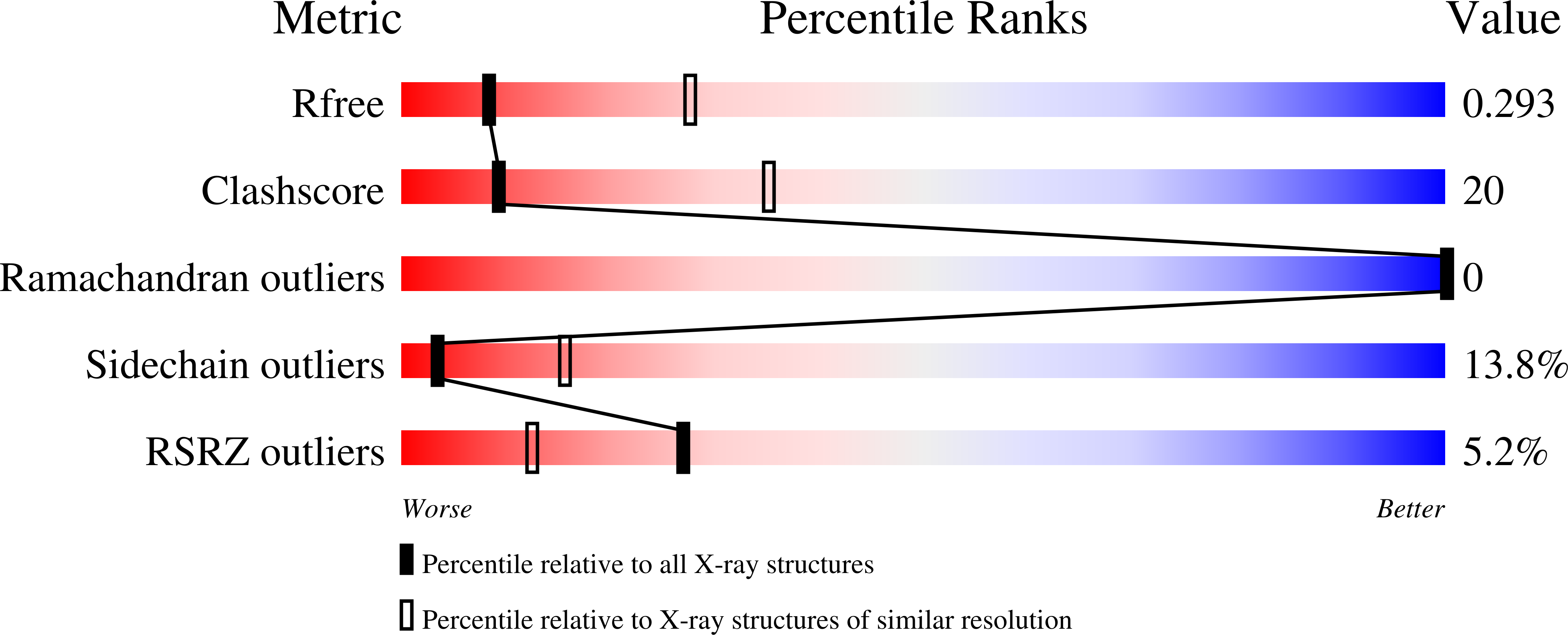
  • R-Value Free: 0.291 
  • R-Value Work: 0.253 
  • R-Value Observed: 0.255 

wwPDB Validation   3D Report Full Report


Ligand Structure Quality Assessment 


This is version 1.5 of the entry. See complete history


Literature

Structure of a eukaryotic SWEET transporter in a homotrimeric complex.

Tao, Y.Cheung, L.S.Li, S.Eom, J.S.Chen, L.Q.Xu, Y.Perry, K.Frommer, W.B.Feng, L.

(2015) Nature 527: 259-263

  • DOI: https://doi.org/10.1038/nature15391
  • Primary Citation of Related Structures:  
    5CTG, 5CTH

  • PubMed Abstract: 

    Eukaryotes rely on efficient distribution of energy and carbon skeletons between organs in the form of sugars. Glucose in animals and sucrose in plants serve as the dominant distribution forms. Cellular sugar uptake and release require vesicular and/or plasma membrane transport proteins. Humans and plants use proteins from three superfamilies for sugar translocation: the major facilitator superfamily (MFS), the sodium solute symporter family (SSF; only in the animal kingdom), and SWEETs. SWEETs carry mono- and disaccharides across vacuolar or plasma membranes. Plant SWEETs play key roles in sugar translocation between compartments, cells, and organs, notably in nectar secretion, phloem loading for long distance translocation, pollen nutrition, and seed filling. Plant SWEETs cause pathogen susceptibility possibly by sugar leakage from infected cells. The vacuolar Arabidopsis thaliana AtSWEET2 sequesters sugars in root vacuoles; loss-of-function mutants show increased susceptibility to Pythium infection. Here we show that its orthologue, the vacuolar glucose transporter OsSWEET2b from rice (Oryza sativa), consists of an asymmetrical pair of triple-helix bundles, connected by an inversion linker transmembrane helix (TM4) to create the translocation pathway. Structural and biochemical analyses show OsSWEET2b in an apparent inward (cytosolic) open state forming homomeric trimers. TM4 tightly interacts with the first triple-helix bundle within a protomer and mediates key contacts among protomers. Structure-guided mutagenesis of the close paralogue SWEET1 from Arabidopsis identified key residues in substrate translocation and protomer crosstalk. Insights into the structure-function relationship of SWEETs are valuable for understanding the transport mechanism of eukaryotic SWEETs and may be useful for engineering sugar flux.


  • Organizational Affiliation

    Department of Molecular and Cellular Physiology, 279 Campus Drive, Stanford University School of Medicine, Stanford, CA 94305, USA.


Macromolecules
Find similar proteins by:  (by identity cutoff)  |  3D Structure
Entity ID: 1
MoleculeChains Sequence LengthOrganismDetailsImage
Bidirectional sugar transporter SWEET2b
A, B, C
224Oryza sativa Japonica GroupMutation(s): 0 
Gene Names: SWEET2BOs01g0700100LOC_Os01g50460OsJ_03146P0047E11.3P0454A11.22
Membrane Entity: Yes 
UniProt
Find proteins for Q5N8J1 (Oryza sativa subsp. japonica)
Explore Q5N8J1 
Go to UniProtKB:  Q5N8J1
Entity Groups  
Sequence Clusters30% Identity50% Identity70% Identity90% Identity95% Identity100% Identity
UniProt GroupQ5N8J1
Sequence Annotations
Expand
  • Reference Sequence
Small Molecules
Ligands 3 Unique
IDChains Name / Formula / InChI Key2D Diagram3D Interactions
PE5
Query on PE5

Download Ideal Coordinates CCD File 
D [auth A],
G [auth B]
3,6,9,12,15,18,21,24-OCTAOXAHEXACOSAN-1-OL
C18 H38 O9
CUDPPTPIUWYGFI-UHFFFAOYSA-N
BNG
Query on BNG

Download Ideal Coordinates CCD File 
E [auth A]nonyl beta-D-glucopyranoside
C15 H30 O6
QFAPUKLCALRPLH-UXXRCYHCSA-N
TRS
Query on TRS

Download Ideal Coordinates CCD File 
F [auth A],
H [auth B],
I [auth C]
2-AMINO-2-HYDROXYMETHYL-PROPANE-1,3-DIOL
C4 H12 N O3
LENZDBCJOHFCAS-UHFFFAOYSA-O
Experimental Data & Validation

Experimental Data

  • Method: X-RAY DIFFRACTION
  • Resolution: 3.10 Å
  • R-Value Free: 0.291 
  • R-Value Work: 0.253 
  • R-Value Observed: 0.255 
  • Space Group: P 1 21 1
Unit Cell:
Length ( Å )Angle ( ˚ )
a = 51.83α = 90
b = 142.24β = 93.36
c = 81.57γ = 90
Software Package:
Software NamePurpose
PHENIXrefinement
HKL-2000data reduction
SCALEPACKdata scaling
PHENIXphasing

Structure Validation

View Full Validation Report


Ligand Structure Quality Assessment 


Entry History & Funding Information

Deposition Data


Funding OrganizationLocationGrant Number
Alfred P. Sloan FoundationUnited States--

Revision History  (Full details and data files)

  • Version 1.0: 2015-10-28
    Type: Initial release
  • Version 1.1: 2015-11-25
    Changes: Database references
  • Version 1.2: 2017-11-01
    Changes: Author supporting evidence, Database references, Derived calculations
  • Version 1.3: 2018-01-17
    Changes: Author supporting evidence
  • Version 1.4: 2020-07-29
    Type: Remediation
    Reason: Carbohydrate remediation
    Changes: Data collection, Derived calculations, Structure summary
  • Version 1.5: 2024-03-06
    Changes: Data collection, Database references, Structure summary