5DDS

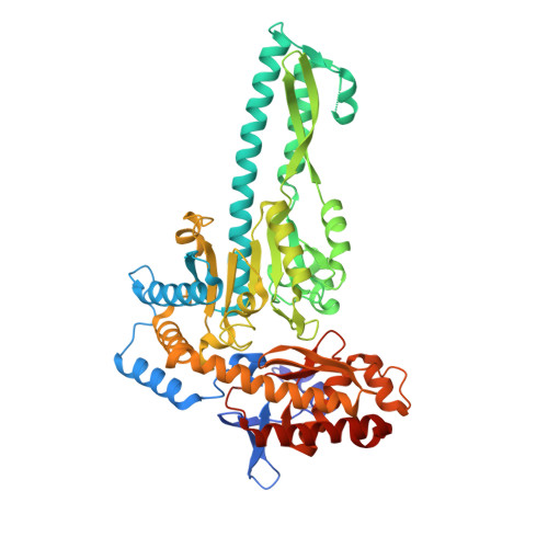
Crystal structure of aminotransferase CrmG from Actinoalloteichus sp. WH1-2216-6 in complex with PLP


Experimental Data Snapshot

  • Method: X-RAY DIFFRACTION
  • Resolution: 2.60 Å
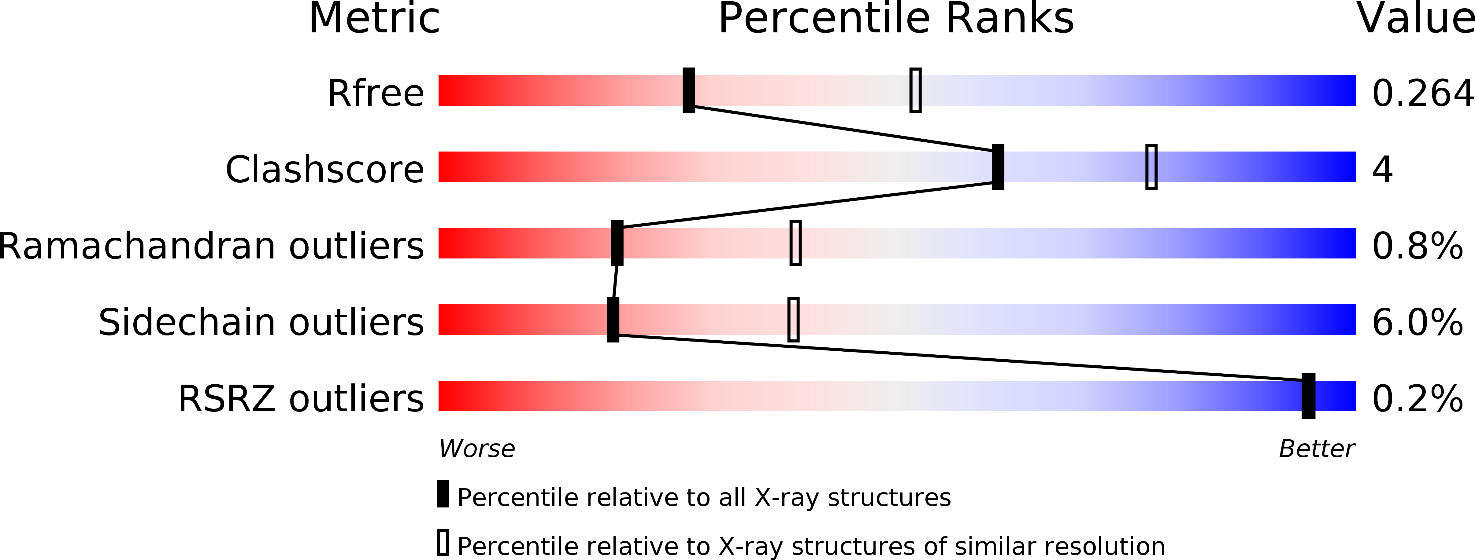
  • R-Value Free: 0.262 
  • R-Value Work: 0.215 
  • R-Value Observed: 0.217 

wwPDB Validation   3D Report Full Report


Ligand Structure Quality Assessment 


This is version 1.0 of the entry. See complete history


Literature

Biochemical and Structural Insights into the Aminotransferase CrmG in Caerulomycin Biosynthesis

Zhu, Y.Xu, J.Mei, X.Feng, Z.Zhang, L.Zhang, Q.Zhang, G.Zhu, W.Liu, J.Zhang, C.

(2016) ACS Chem Biol 11: 943-952

  • DOI: https://doi.org/10.1021/acschembio.5b00984
  • Primary Citation of Related Structures:  
    5DDS, 5DDU, 5DDW

  • PubMed Abstract: 

    Caerulomycin A (CRM A 1) belongs to a family of natural products containing a 2,2'-bipyridyl ring core structure and is currently under development as a potent novel immunosuppressive agent. Herein, we report the functional characterization, kinetic analysis, substrate specificity, and structure insights of an aminotransferase CrmG in 1 biosynthesis. The aminotransferase CrmG was confirmed to catalyze a key transamination reaction to convert an aldehyde group to an amino group in the 1 biosynthetic pathway, preferring l-glutamate and l-glutamine as the amino donor substrates. The crystal structures of CrmG in complex with the cofactor 5'-pyridoxal phosphate (PLP) or 5'-pyridoxamine phosphate (PMP) or the acceptor substrate were determined to adopt a canonical fold-type I of PLP-dependent enzymes with a unique small additional domain. The structure guided site-directed mutagenesis identified key amino acid residues for substrate binding and catalytic activities, thus providing insights into the transamination mechanism of CrmG.


  • Organizational Affiliation

    CAS Key Laboratory of Tropical Marine Bio-resources and Ecology, Guangdong Key Laboratory of Marine Materia Medica, RNAM Center for Marine Microbiology, South China Sea Institute of Oceanology, Chinese Academy of Sciences , 164 West Xingang Road, Guangzhou 510301, China.


Macromolecules
Find similar proteins by:  (by identity cutoff)  |  3D Structure
Entity ID: 1
MoleculeChains Sequence LengthOrganismDetailsImage
CrmG
A, B, C, D
523Actinoalloteichus sp. WH1-2216-6Mutation(s): 0 
UniProt
Find proteins for H8Y6N2 (Actinoalloteichus sp. WH1-2216-6)
Explore H8Y6N2 
Go to UniProtKB:  H8Y6N2
Entity Groups  
Sequence Clusters30% Identity50% Identity70% Identity90% Identity95% Identity100% Identity
UniProt GroupH8Y6N2
Sequence Annotations
Expand
  • Reference Sequence
Small Molecules
Ligands 3 Unique
IDChains Name / Formula / InChI Key2D Diagram3D Interactions
PLP
Query on PLP

Download Ideal Coordinates CCD File 
E [auth A],
G [auth B],
I [auth C],
J [auth D]
PYRIDOXAL-5'-PHOSPHATE
C8 H10 N O6 P
NGVDGCNFYWLIFO-UHFFFAOYSA-N
GOL
Query on GOL

Download Ideal Coordinates CCD File 
F [auth A],
H [auth B]
GLYCEROL
C3 H8 O3
PEDCQBHIVMGVHV-UHFFFAOYSA-N
ACY
Query on ACY

Download Ideal Coordinates CCD File 
K [auth D]ACETIC ACID
C2 H4 O2
QTBSBXVTEAMEQO-UHFFFAOYSA-N
Experimental Data & Validation

Experimental Data

  • Method: X-RAY DIFFRACTION
  • Resolution: 2.60 Å
  • R-Value Free: 0.262 
  • R-Value Work: 0.215 
  • R-Value Observed: 0.217 
  • Space Group: P 1
Unit Cell:
Length ( Å )Angle ( ˚ )
a = 84.078α = 106.71
b = 84.21β = 109.16
c = 88.949γ = 95.03
Software Package:
Software NamePurpose
MOSFLMdata collection
Aimlessdata scaling
REFMACrefinement
PDB_EXTRACTdata extraction
Aimlessdata reduction
PHENIXphasing

Structure Validation

View Full Validation Report


Ligand Structure Quality Assessment 


Entry History & Funding Information

Deposition Data


Funding OrganizationLocationGrant Number
National Science Foundation of ChinaChina31500638
Natural Science Foundation of Guangdong ProvinceChina2014A030310356

Revision History  (Full details and data files)

  • Version 1.0: 2016-08-10
    Type: Initial release