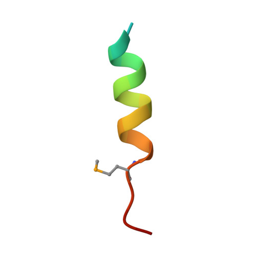
5DI9

Crystal Structure of hRio2 NES Reverse Mutant Peptide in complex with CRM1-Ran-RanBP1


Experimental Data Snapshot

  • Method: X-RAY DIFFRACTION
  • Resolution: 2.28 Å
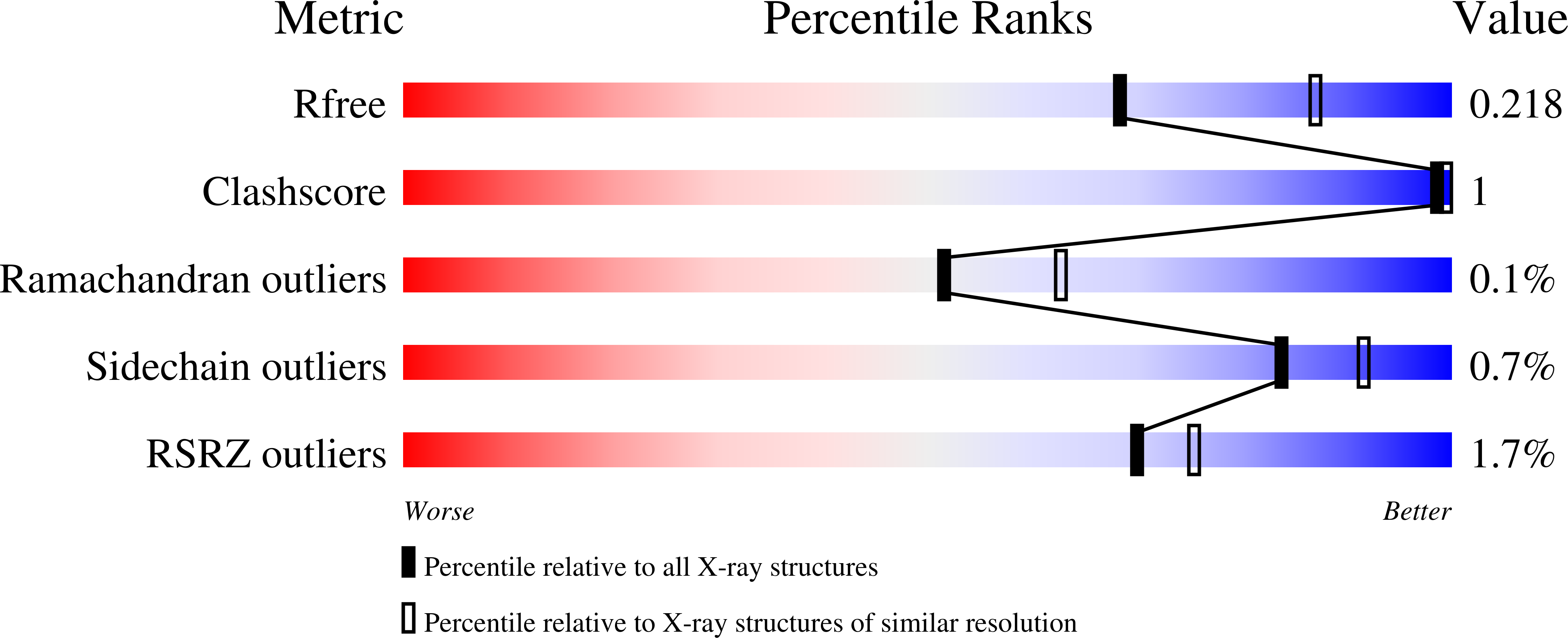
  • R-Value Free: 0.211 
  • R-Value Work: 0.168 
  • R-Value Observed: 0.169 

Starting Model: experimental
View more details

wwPDB Validation   3D Report Full Report


Ligand Structure Quality Assessment 


This is version 1.8 of the entry. See complete history


Literature

Structural determinants of nuclear export signal orientation in binding to exportin CRM1.

Fung, H.Y.Fu, S.C.Brautigam, C.A.Chook, Y.M.

(2015) Elife 4

  • DOI: https://doi.org/10.7554/eLife.10034
  • Primary Citation of Related Structures:  
    5DH9, 5DHA, 5DHF, 5DI9, 5DIF

  • PubMed Abstract: 

    The Chromosome Region of Maintenance 1 (CRM1) protein mediates nuclear export of hundreds of proteins through recognition of their nuclear export signals (NESs), which are highly variable in sequence and structure. The plasticity of the CRM1-NES interaction is not well understood, as there are many NES sequences that seem incompatible with structures of the NES-bound CRM1 groove. Crystal structures of CRM1 bound to two different NESs with unusual sequences showed the NES peptides binding the CRM1 groove in the opposite orientation (minus) to that of previously studied NESs (plus). Comparison of minus and plus NESs identified structural and sequence determinants for NES orientation. The binding of NESs to CRM1 in both orientations results in a large expansion in NES consensus patterns and therefore a corresponding expansion of potential NESs in the proteome.


  • Organizational Affiliation

    Department of Pharmacology, University of Texas Southwestern Medical Center, Dallas, United States.


Macromolecules
Find similar proteins by:  (by identity cutoff)  |  3D Structure
Entity ID: 1
MoleculeChains Sequence LengthOrganismDetailsImage
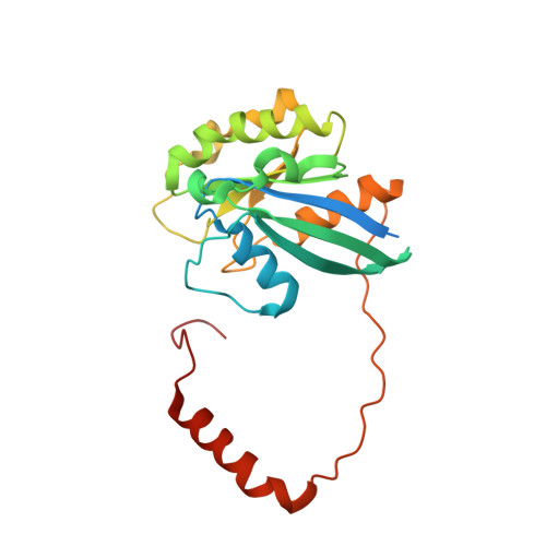
GTP-binding nuclear protein Ran237Homo sapiensMutation(s): 0 
Gene Names: RANARA24OK/SW-cl.81
EC: 3.6.5
UniProt & NIH Common Fund Data Resources
Find proteins for P62826 (Homo sapiens)
Explore P62826 
Go to UniProtKB:  P62826
PHAROS:  P62826
GTEx:  ENSG00000132341 
Entity Groups  
Sequence Clusters30% Identity50% Identity70% Identity90% Identity95% Identity100% Identity
UniProt GroupP62826
Sequence Annotations
Expand
  • Reference Sequence
Find similar proteins by:  (by identity cutoff)  |  3D Structure
Entity ID: 2
MoleculeChains Sequence LengthOrganismDetailsImage
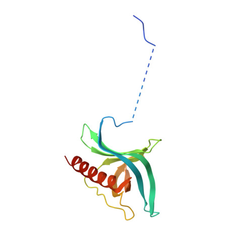
Ran-specific GTPase-activating protein 1143Saccharomyces cerevisiae S288CMutation(s): 0 
Gene Names: YRB1CST20HTN1SFO1YDR002WYD8119.08
UniProt
Find proteins for P41920 (Saccharomyces cerevisiae (strain ATCC 204508 / S288c))
Explore P41920 
Go to UniProtKB:  P41920
Entity Groups  
Sequence Clusters30% Identity50% Identity70% Identity90% Identity95% Identity100% Identity
UniProt GroupP41920
Sequence Annotations
Expand
  • Reference Sequence
Find similar proteins by:  (by identity cutoff)  |  3D Structure
Entity ID: 3
MoleculeChains Sequence LengthOrganismDetailsImage
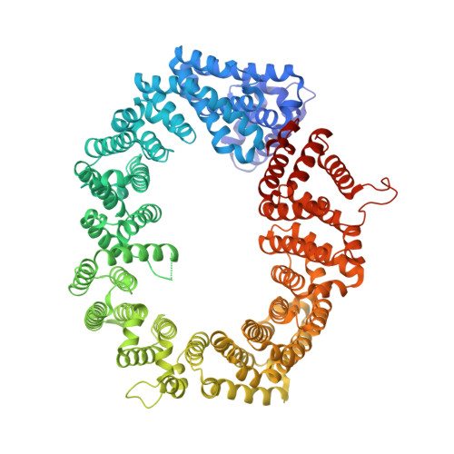
Exportin-11,024Saccharomyces cerevisiae S288CMutation(s): 6 
Gene Names: CRM1KAP124XPO1YGR218WG8514
UniProt
Find proteins for P30822 (Saccharomyces cerevisiae (strain ATCC 204508 / S288c))
Explore P30822 
Go to UniProtKB:  P30822
Entity Groups  
Sequence Clusters30% Identity50% Identity70% Identity90% Identity95% Identity100% Identity
UniProt GroupP30822
Sequence Annotations
Expand
  • Reference Sequence

Find similar proteins by:  Sequence   |   3D Structure  

Entity ID: 4
MoleculeChains Sequence LengthOrganismDetailsImage
Engineered Nuclear Export Signal Peptide (hRio2 NES reverse mutant)21Homo sapiensMutation(s): 0 
Gene Names: CPEB4
Entity Groups  
Sequence Clusters30% Identity50% Identity70% Identity90% Identity95% Identity100% Identity
Sequence Annotations
Expand
  • Reference Sequence
Small Molecules
Ligands 5 Unique
IDChains Name / Formula / InChI Key2D Diagram3D Interactions
GNP
Query on GNP

Download Ideal Coordinates CCD File 
E [auth A]PHOSPHOAMINOPHOSPHONIC ACID-GUANYLATE ESTER
C10 H17 N6 O13 P3
UQABYHGXWYXDTK-UUOKFMHZSA-N
GOL
Query on GOL

Download Ideal Coordinates CCD File 
G [auth A],
J [auth C],
K [auth C],
L [auth C]
GLYCEROL
C3 H8 O3
PEDCQBHIVMGVHV-UHFFFAOYSA-N
ZN
Query on ZN

Download Ideal Coordinates CCD File 
H [auth C]ZINC ION
Zn
PTFCDOFLOPIGGS-UHFFFAOYSA-N
CL
Query on CL

Download Ideal Coordinates CCD File 
I [auth C]CHLORIDE ION
Cl
VEXZGXHMUGYJMC-UHFFFAOYSA-M
MG
Query on MG

Download Ideal Coordinates CCD File 
F [auth A]MAGNESIUM ION
Mg
JLVVSXFLKOJNIY-UHFFFAOYSA-N
Modified Residues  1 Unique
IDChains TypeFormula2D DiagramParent
MSE
Query on MSE
D
L-PEPTIDE LINKINGC5 H11 N O2 SeMET
Experimental Data & Validation

Experimental Data

  • Method: X-RAY DIFFRACTION
  • Resolution: 2.28 Å
  • R-Value Free: 0.211 
  • R-Value Work: 0.168 
  • R-Value Observed: 0.169 
  • Space Group: P 43 21 2
Unit Cell:
Length ( Å )Angle ( ˚ )
a = 106.692α = 90
b = 106.692β = 90
c = 304.497γ = 90
Software Package:
Software NamePurpose
PHENIXrefinement
HKL-2000data scaling
PDB_EXTRACTdata extraction
Cootmodel building

Structure Validation

View Full Validation Report


Ligand Structure Quality Assessment 


Entry History & Funding Information

Deposition Data


Funding OrganizationLocationGrant Number
Croucher FoundationHong KongStudent Scholarship
National Institutes of Health/National Institute of General Medical Sciences (NIH/NIGMS)United StatesGM069909
Cancer Prevention and Research Institute of Texas (CPRIT)United StatesRP120352
University of Texas SouthwesternUnited StatesEndowed Scholars Program
Welch FoundationUnited StatesI-1532
Leukemia & Lymphoma SocietyUnited StatesScholar Award

Revision History  (Full details and data files)

  • Version 1.0: 2015-09-16
    Type: Initial release
  • Version 1.1: 2015-10-21
    Changes: Database references
  • Version 1.2: 2017-09-20
    Changes: Author supporting evidence, Derived calculations
  • Version 1.3: 2017-09-27
    Changes: Author supporting evidence
  • Version 1.4: 2019-04-24
    Changes: Author supporting evidence, Data collection
  • Version 1.5: 2019-12-25
    Changes: Author supporting evidence
  • Version 1.6: 2023-09-27
    Changes: Data collection, Database references, Derived calculations, Refinement description
  • Version 1.7: 2023-11-15
    Changes: Data collection
  • Version 1.8: 2024-11-20
    Changes: Structure summary