5DJT

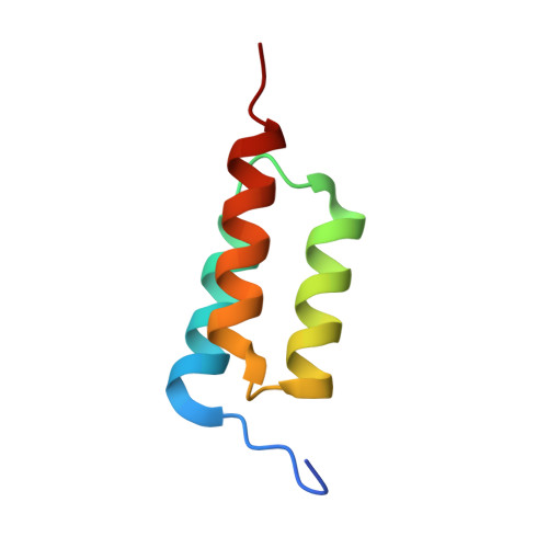
Crystal structure of LOV2 (C450A) domain in complex with Zdk2


Experimental Data Snapshot

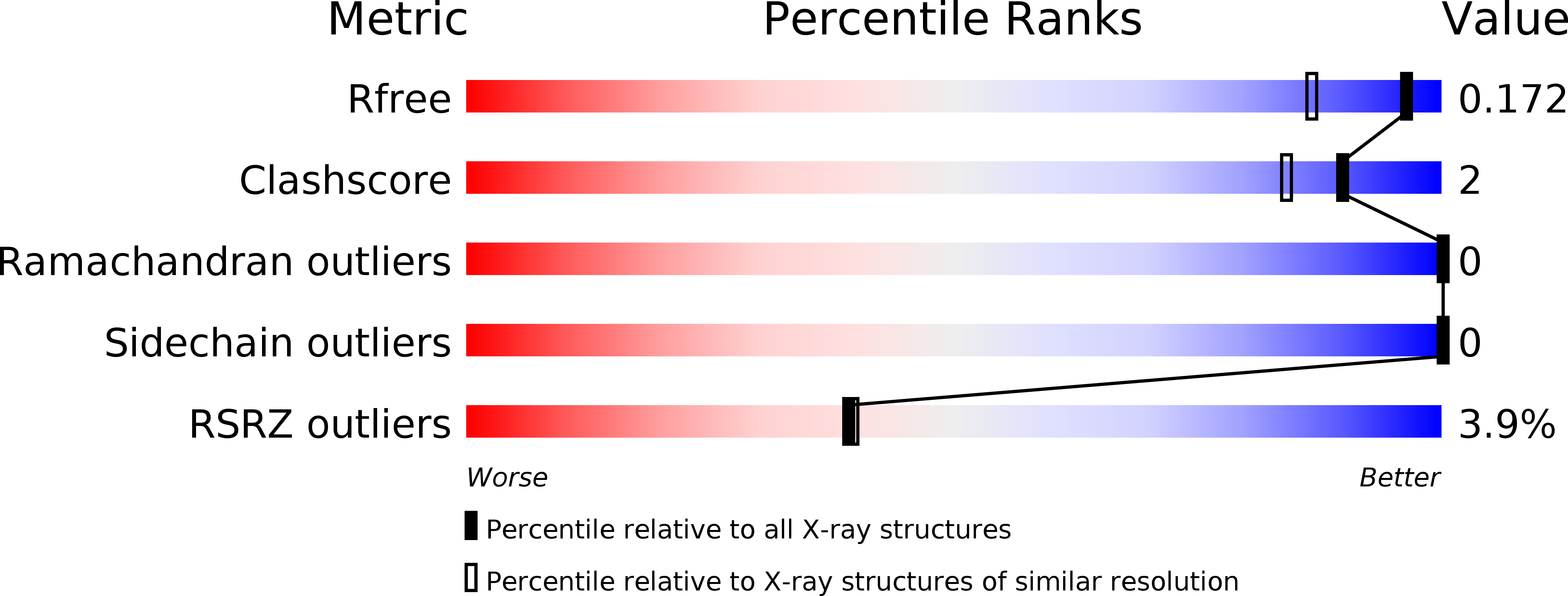
  • Method: X-RAY DIFFRACTION
  • Resolution: 1.40 Å
  • R-Value Free: 0.171 
  • R-Value Work: 0.146 
  • R-Value Observed: 0.147 

Starting Models: experimental
View more details

wwPDB Validation   3D Report Full Report


Ligand Structure Quality Assessment 


This is version 1.3 of the entry. See complete history


Literature

LOVTRAP: an optogenetic system for photoinduced protein dissociation.

Wang, H.Vilela, M.Winkler, A.Tarnawski, M.Schlichting, I.Yumerefendi, H.Kuhlman, B.Liu, R.Danuser, G.Hahn, K.M.

(2016) Nat Methods 13: 755-758

  • DOI: https://doi.org/10.1038/nmeth.3926
  • Primary Citation of Related Structures:  
    5DJT, 5DJU, 5EFW

  • PubMed Abstract: 

    LOVTRAP is an optogenetic approach for reversible light-induced protein dissociation using protein A fragments that bind to the LOV domain only in the dark, with tunable kinetics and a >150-fold change in the dissociation constant (Kd). By reversibly sequestering proteins at mitochondria, we precisely modulated the proteins' access to the cell edge, demonstrating a naturally occurring 3-mHz cell-edge oscillation driven by interactions of Vav2, Rac1, and PI3K proteins.


  • Organizational Affiliation

    Department of Pharmacology, University of North Carolina at Chapel Hill, Chapel Hill, North Carolina, USA.


Macromolecules
Find similar proteins by:  (by identity cutoff)  |  3D Structure
Entity ID: 1
MoleculeChains Sequence LengthOrganismDetailsImage
NPH1-2145Avena sativaMutation(s): 0 
Gene Names: NPH1-2
EC: 2.7.11.1
UniProt
Find proteins for O49004 (Avena sativa)
Explore O49004 
Go to UniProtKB:  O49004
Entity Groups  
Sequence Clusters30% Identity50% Identity70% Identity90% Identity95% Identity100% Identity
UniProt GroupO49004
Sequence Annotations
Expand
  • Reference Sequence
Find similar proteins by:  (by identity cutoff)  |  3D Structure
Entity ID: 2
MoleculeChains Sequence LengthOrganismDetailsImage
Engineered protein, Zdk2 affibody61Staphylococcus aureusMutation(s): 0 
Entity Groups  
Sequence Clusters30% Identity50% Identity70% Identity90% Identity95% Identity100% Identity
Sequence Annotations
Expand
  • Reference Sequence
Experimental Data & Validation

Experimental Data

  • Method: X-RAY DIFFRACTION
  • Resolution: 1.40 Å
  • R-Value Free: 0.171 
  • R-Value Work: 0.146 
  • R-Value Observed: 0.147 
  • Space Group: I 4
Unit Cell:
Length ( Å )Angle ( ˚ )
a = 110.14α = 90
b = 110.14β = 90
c = 37.51γ = 90
Software Package:
Software NamePurpose
PHENIXrefinement
XDSdata reduction
XSCALEdata scaling
PHASERphasing

Structure Validation

View Full Validation Report


Ligand Structure Quality Assessment 


Entry History 

Deposition Data

Revision History  (Full details and data files)

  • Version 1.0: 2016-07-20
    Type: Initial release
  • Version 1.1: 2016-07-27
    Changes: Database references
  • Version 1.2: 2016-09-07
    Changes: Database references
  • Version 1.3: 2024-01-10
    Changes: Data collection, Database references, Refinement description