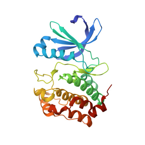
Allosteric modulation of AURKA kinase activity by a small-molecule inhibitor of its protein-protein interaction with TPX2.
Janecek, M., Rossmann, M., Sharma, P., Emery, A., Huggins, D.J., Stockwell, S.R., Stokes, J.E., Tan, Y.S., Almeida, E.G., Hardwick, B., Narvaez, A.J., Hyvonen, M., Spring, D.R., McKenzie, G.J., Venkitaraman, A.R.(2016) Sci Rep 6: 28528-28528
- PubMed: 27339427
- DOI: https://doi.org/10.1038/srep28528
- Primary Citation of Related Structures:
5DN3, 5DNR, 5DOS, 5DPV, 5DR2, 5DR6, 5DR9, 5DRD, 5DT0, 5DT3, 5DT4 - PubMed Abstract:
The essential mitotic kinase Aurora A (AURKA) is controlled during cell cycle progression via two distinct mechanisms. Following activation loop autophosphorylation early in mitosis when it localizes to centrosomes, AURKA is allosterically activated on the mitotic spindle via binding to the microtubule-associated protein, TPX2. Here, we report the discovery of AurkinA, a novel chemical inhibitor of the AURKA-TPX2 interaction, which acts via an unexpected structural mechanism to inhibit AURKA activity and mitotic localization. In crystal structures, AurkinA binds to a hydrophobic pocket (the 'Y pocket') that normally accommodates a conserved Tyr-Ser-Tyr motif from TPX2, blocking the AURKA-TPX2 interaction. AurkinA binding to the Y- pocket induces structural changes in AURKA that inhibit catalytic activity in vitro and in cells, without affecting ATP binding to the active site, defining a novel mechanism of allosteric inhibition. Consistent with this mechanism, cells exposed to AurkinA mislocalise AURKA from mitotic spindle microtubules. Thus, our findings provide fresh insight into the catalytic mechanism of AURKA, and identify a key structural feature as the target for a new class of dual-mode AURKA inhibitors, with implications for the chemical biology and selective therapeutic targeting of structurally related kinases.
Organizational Affiliation:
MRC Cancer Unit, University of Cambridge, Hills Road, Cambridge CB2 0XZ, United Kingdom.


