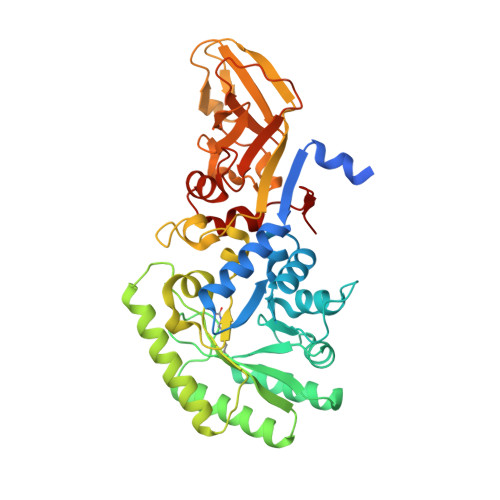
5FAC

Alanine Racemase from Streptomyces coelicolor A3(2)


Experimental Data Snapshot

  • Method: X-RAY DIFFRACTION
  • Resolution: 2.80 Å
  • R-Value Free: 0.259 
  • R-Value Work: 0.208 
  • R-Value Observed: 0.211 

Starting Model: experimental
View more details

wwPDB Validation   3D Report Full Report


Ligand Structure Quality Assessment 


This is version 1.3 of the entry. See complete history


Literature

Structural and functional characterization of the alanine racemase from Streptomyces coelicolor A3(2).

Tassoni, R.van der Aart, L.T.Ubbink, M.van Wezel, G.P.Pannu, N.S.

(2017) Biochem Biophys Res Commun 483: 122-128

  • DOI: https://doi.org/10.1016/j.bbrc.2016.12.183
  • Primary Citation of Related Structures:  
    5FAC, 5FAG, 5FAJ

  • PubMed Abstract: 

    The conversion of l-alanine (L-Ala) into d-alanine (D-Ala) in bacteria is performed by pyridoxal phosphate-dependent enzymes called alanine racemases. D-Ala is an essential component of the bacterial peptidoglycan and hence required for survival. The Gram-positive bacterium Streptomyces coelicolor has at least one alanine racemase encoded by alr. Here, we describe an alr deletion mutant of S. coelicolor which depends on D-Ala for growth and shows increased sensitivity to the antibiotic d-cycloserine (DCS). The crystal structure of the alanine racemase (Alr) was solved with and without the inhibitors DCS or propionate, at 1.64 Å and 1.51 Å resolution, respectively. The crystal structures revealed that Alr is a homodimer with residues from both monomers contributing to the active site. The dimeric state of the enzyme in solution was confirmed by gel filtration chromatography, with and without L-Ala or d-cycloserine. The activity of the enzyme was 66 ± 3 U mg -1 for the racemization of L- to D-Ala, and 104 ± 7 U mg -1 for the opposite direction. Comparison of Alr from S. coelicolor with orthologous enzymes from other bacteria, including the closely related d-cycloserine-resistant Alr from S. lavendulae, strongly suggests that structural features such as the hinge angle or the surface area between the monomers do not contribute to d-cycloserine resistance, and the molecular basis for resistance therefore remains elusive.


  • Organizational Affiliation

    Leiden Institute of Chemistry, Leiden University, Gorlaeus Laboratories, Einsteinweg 55, 2333 CC Leiden, The Netherlands.


Macromolecules
Find similar proteins by:  (by identity cutoff)  |  3D Structure
Entity ID: 1
MoleculeChains Sequence LengthOrganismDetailsImage
Alanine racemase
A, B, C, D
410Streptomyces coelicolor A3(2)Mutation(s): 0 
Gene Names: alrSCO4745SC6G4.23
EC: 5.1.1.1
UniProt
Find proteins for O86786 (Streptomyces coelicolor (strain ATCC BAA-471 / A3(2) / M145))
Explore O86786 
Go to UniProtKB:  O86786
Entity Groups  
Sequence Clusters30% Identity50% Identity70% Identity90% Identity95% Identity100% Identity
UniProt GroupO86786
Sequence Annotations
Expand
  • Reference Sequence
Small Molecules
Ligands 2 Unique
IDChains Name / Formula / InChI Key2D Diagram3D Interactions
PLP
Query on PLP

Download Ideal Coordinates CCD File 
E [auth A],
G [auth B],
I [auth C],
K [auth D]
PYRIDOXAL-5'-PHOSPHATE
C8 H10 N O6 P
NGVDGCNFYWLIFO-UHFFFAOYSA-N
CL
Query on CL

Download Ideal Coordinates CCD File 
F [auth A],
H [auth B],
J [auth C],
L [auth D]
CHLORIDE ION
Cl
VEXZGXHMUGYJMC-UHFFFAOYSA-M
Modified Residues  1 Unique
IDChains TypeFormula2D DiagramParent
KCX
Query on KCX
A, B, C, D
L-PEPTIDE LINKINGC7 H14 N2 O4LYS
Experimental Data & Validation

Experimental Data

  • Method: X-RAY DIFFRACTION
  • Resolution: 2.80 Å
  • R-Value Free: 0.259 
  • R-Value Work: 0.208 
  • R-Value Observed: 0.211 
  • Space Group: P 1 21 1
Unit Cell:
Length ( Å )Angle ( ˚ )
a = 78.96α = 90
b = 87.17β = 102.26
c = 109.626γ = 90
Software Package:
Software NamePurpose
REFMACrefinement
XDSdata reduction
Aimlessdata scaling
MOLREPphasing

Structure Validation

View Full Validation Report


Ligand Structure Quality Assessment 


Entry History 

Deposition Data

Revision History  (Full details and data files)

  • Version 1.0: 2016-12-21
    Type: Initial release
  • Version 1.1: 2017-01-25
    Changes: Database references
  • Version 1.2: 2017-02-01
    Changes: Database references
  • Version 1.3: 2024-01-10
    Changes: Data collection, Database references, Refinement description