Carbohydrate-binding domain of the POMGnT1 stem region modulates O-mannosylation sites of alpha-dystroglycan
Kuwabara, N., Manya, H., Yamada, T., Tateno, H., Kanagawa, M., Kobayashi, K., Akasaka-Manya, K., Hirose, Y., Mizuno, M., Ikeguchi, M., Toda, T., Hirabayashi, J., Senda, T., Endo, T., Kato, R.(2016) Proc Natl Acad Sci U S A 113: 9280-9285
- PubMed: 27493216
- DOI: https://doi.org/10.1073/pnas.1525545113
- Primary Citation of Related Structures:
5GGF, 5GGG, 5GGI, 5GGJ, 5GGK, 5GGL, 5GGN, 5GGO, 5GGP - PubMed Abstract:
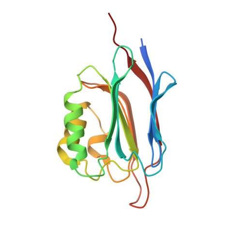
The dystrophin glycoprotein complex, which connects the cell membrane to the basement membrane, is essential for a variety of biological events, including maintenance of muscle integrity. An O-mannose-type GalNAc-β1,3-GlcNAc-β1,4-(phosphate-6)-Man structure of α-dystroglycan (α-DG), a subunit of the complex that is anchored to the cell membrane, interacts directly with laminin in the basement membrane. Reduced glycosylation of α-DG is linked to some types of inherited muscular dystrophy; consistent with this relationship, many disease-related mutations have been detected in genes involved in O-mannosyl glycan synthesis. Defects in protein O-linked mannose β1,2-N-acetylglucosaminyltransferase 1 (POMGnT1), a glycosyltransferase that participates in the formation of GlcNAc-β1,2-Man glycan, are causally related to muscle-eye-brain disease (MEB), a congenital muscular dystrophy, although the role of POMGnT1 in postphosphoryl modification of GalNAc-β1,3-GlcNAc-β1,4-(phosphate-6)-Man glycan remains elusive. Our crystal structures of POMGnT1 agreed with our previous results showing that the catalytic domain recognizes substrate O-mannosylated proteins via hydrophobic interactions with little sequence specificity. Unexpectedly, we found that the stem domain recognizes the β-linked GlcNAc of O-mannosyl glycan, an enzymatic product of POMGnT1. This interaction may recruit POMGnT1 to a specific site of α-DG to promote GlcNAc-β1,2-Man clustering and also may recruit other enzymes that interact with POMGnT1, e.g., fukutin, which is required for further modification of the GalNAc-β1,3-GlcNAc-β1,4-(phosphate-6)-Man glycan. On the basis of our findings, we propose a mechanism for the deficiency in postphosphoryl modification of the glycan observed in POMGnT1-KO mice and MEB patients.
Organizational Affiliation:
Photon Factory, Structural Biology Research Center, Institute of Materials Structure Science, High Energy Accelerator Research Organization, Tsukuba, Ibaraki 305-0801, Japan;


