5HPZ

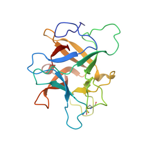
type II water soluble Chl binding proteins


Experimental Data Snapshot

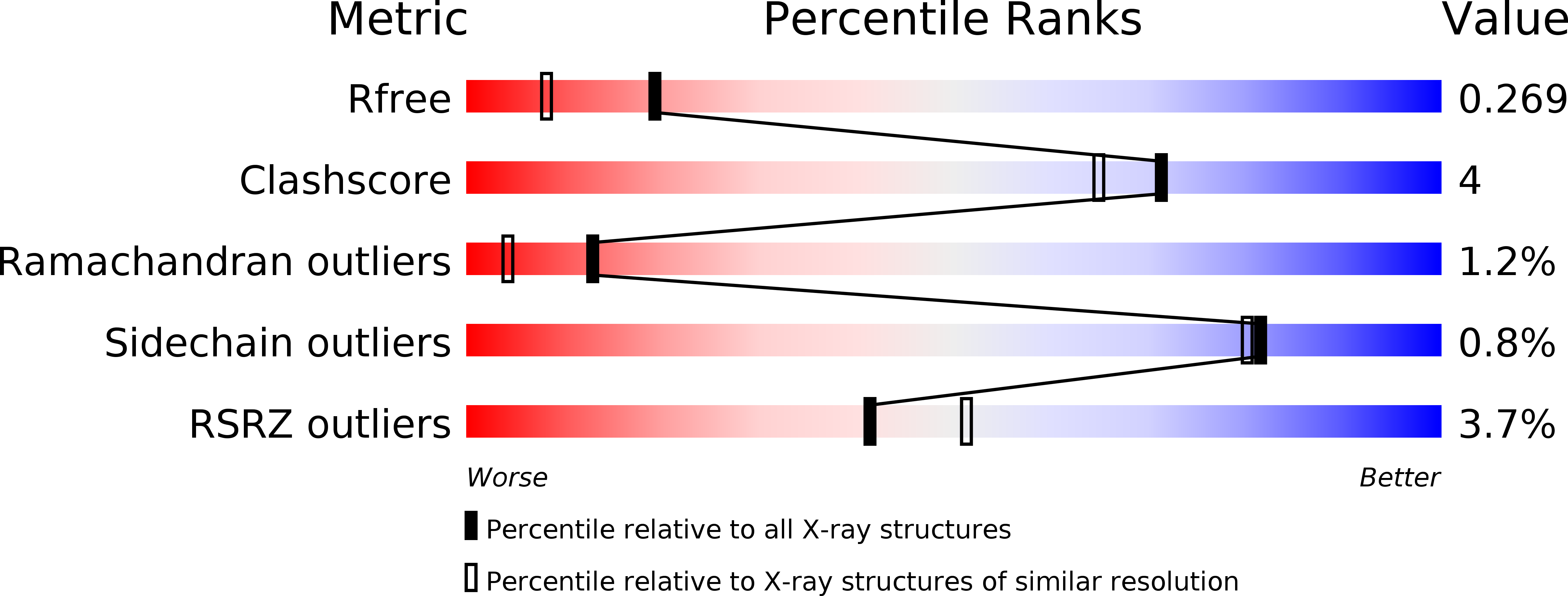
  • Method: X-RAY DIFFRACTION
  • Resolution: 1.96 Å
  • R-Value Free: 0.266 
  • R-Value Work: 0.210 
  • R-Value Observed: 0.213 

wwPDB Validation   3D Report Full Report


Ligand Structure Quality Assessment 


This is version 1.4 of the entry. See complete history


Literature

Fine Tuning of Chlorophyll Spectra by Protein-Induced Ring Deformation.

Bednarczyk, D.Dym, O.Prabahar, V.Peleg, Y.Pike, D.H.Noy, D.

(2016) Angew Chem Int Ed Engl 55: 6901-6905

  • DOI: https://doi.org/10.1002/anie.201512001
  • Primary Citation of Related Structures:  
    5HPZ

  • PubMed Abstract: 

    The ability to tune the light-absorption properties of chlorophylls by their protein environment is the key to the robustness and high efficiency of photosynthetic light-harvesting proteins. Unfortunately, the intricacy of the natural complexes makes it very difficult to identify and isolate specific protein-pigment interactions that underlie the spectral-tuning mechanisms. Herein we identify and demonstrate the tuning mechanism of chlorophyll spectra in type II water-soluble chlorophyll binding proteins from Brassicaceae (WSCPs). By comparing the molecular structures of two natural WSCPs we correlate a shift in the chlorophyll red absorption band with deformation of its tetrapyrrole macrocycle that is induced by changing the position of a nearby tryptophan residue. We show by a set of reciprocal point mutations that this change accounts for up to 2/3 of the observed spectral shift between the two natural variants.


  • Organizational Affiliation

    Department of Biological Chemistry, Weizmann Institute of Science, Rehovot, Israel.


Macromolecules
Find similar proteins by:  (by identity cutoff)  |  3D Structure
Entity ID: 1
MoleculeChains Sequence LengthOrganismDetailsImage
Water-soluble chlorophyll protein
A, B
179BrassicaceaeMutation(s): 0 
Gene Names: WSCP1R
UniProt
Find proteins for Q8H0F0 (Brassica oleracea var. viridis)
Explore Q8H0F0 
Go to UniProtKB:  Q8H0F0
Entity Groups  
Sequence Clusters30% Identity50% Identity70% Identity90% Identity95% Identity100% Identity
UniProt GroupQ8H0F0
Sequence Annotations
Expand
  • Reference Sequence
Experimental Data & Validation

Experimental Data

  • Method: X-RAY DIFFRACTION
  • Resolution: 1.96 Å
  • R-Value Free: 0.266 
  • R-Value Work: 0.210 
  • R-Value Observed: 0.213 
  • Space Group: P 2 21 21
Unit Cell:
Length ( Å )Angle ( ˚ )
a = 38.92α = 90
b = 95.479β = 90
c = 98.61γ = 90
Software Package:
Software NamePurpose
REFMACrefinement
MOSFLMdata reduction
PHASERphasing

Structure Validation

View Full Validation Report


Ligand Structure Quality Assessment 


Entry History 

Deposition Data

Revision History  (Full details and data files)

  • Version 1.0: 2016-05-04
    Type: Initial release
  • Version 1.1: 2016-05-18
    Changes: Derived calculations
  • Version 1.2: 2016-06-08
    Changes: Database references
  • Version 1.3: 2019-05-29
    Changes: Data collection, Derived calculations, Structure summary
  • Version 1.4: 2024-10-23
    Changes: Data collection, Database references, Structure summary