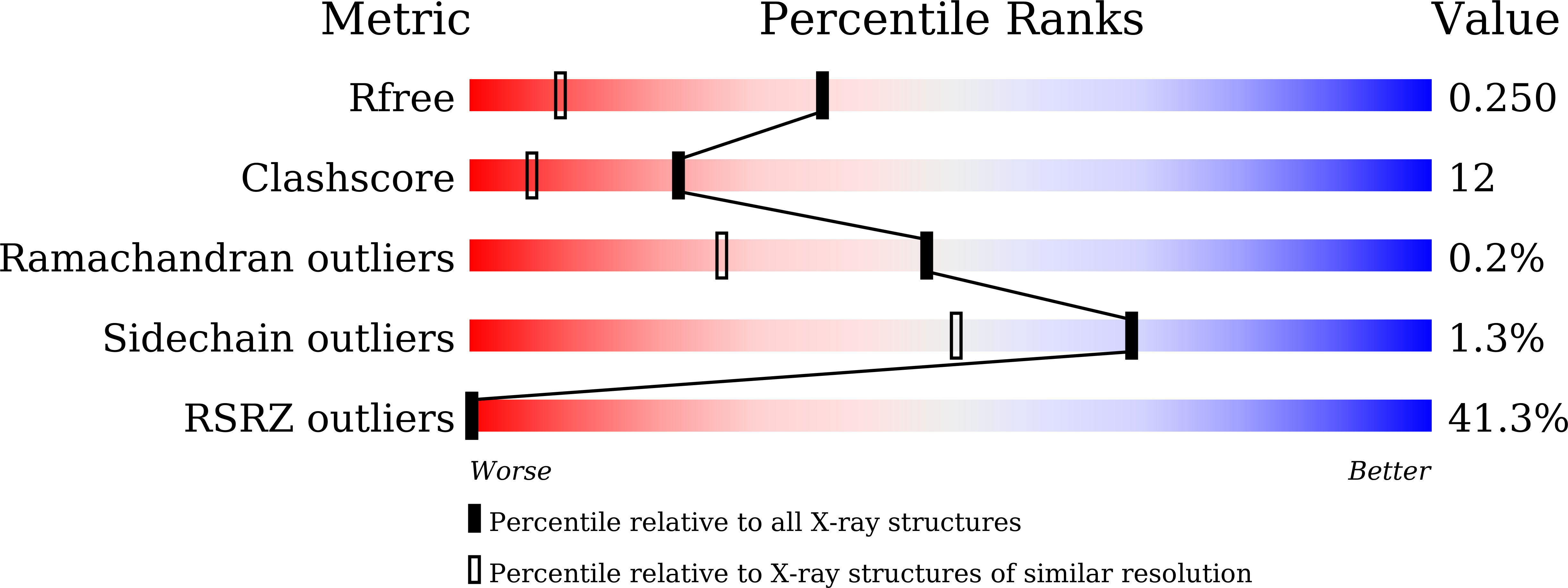
5HRA

Crystal structure of an aspartate/glutamate racemase in complex with D-aspartate


Experimental Data Snapshot

  • Method: X-RAY DIFFRACTION
  • Resolution: 1.60 Å
  • R-Value Free: 0.250 
  • R-Value Work: 0.233 
  • R-Value Observed: 0.234 

Starting Model: experimental
View more details

wwPDB Validation   3D Report Full Report


This is version 1.2 of the entry. See complete history


Literature

Crystal structure and molecular mechanism of an aspartate/glutamate racemase from Escherichia coli O157

Liu, X.Gao, F.Ma, Y.Liu, S.Cui, Y.Yuan, Z.Kang, X.

(2016) FEBS Lett 590: 1262-1269

  • DOI: https://doi.org/10.1002/1873-3468.12148
  • Primary Citation of Related Structures:  
    5HQT, 5HRA, 5HRC

  • PubMed Abstract: 

    EcL-DER, the aspartate/glutamate racemase from the pathogen Escherichia coli O157, exhibits racemase activity for l-aspartate and l-glutamate. This study reports the crystal structures of apo-EcL-DER, the EcL-DER-l-aspartate and the EcL-DER-d-aspartate complexes. The EcL-DER structure contains two domains, forming pseudo-mirror symmetry in the active site. A unique catalytic pair consisting of Thr(83) and Cys(197) exists in the active site. The characteristic conformations of l-Asp and d-Asp in the active site provide a straight structural evidence for the racemization mechanism of EcL-DER. In addition, the diversity of catalytic pairs implies that PLP-independent amino acid racemases adopt various catalytic mechanisms and are classified into different subgroups.


  • Organizational Affiliation

    College of Life Sciences, Hebei University, Baoding, China.


Macromolecules
Find similar proteins by:  (by identity cutoff)  |  3D Structure
Entity ID: 1
MoleculeChains Sequence LengthOrganismDetailsImage
aspartate/glutamate racemase
A, B
235Escherichia coli O157:H7 str. SS52Mutation(s): 0 
Gene Names: ygeASS52_3985
Entity Groups  
Sequence Clusters30% Identity50% Identity70% Identity90% Identity95% Identity100% Identity
Sequence Annotations
Expand
  • Reference Sequence
Experimental Data & Validation

Experimental Data

  • Method: X-RAY DIFFRACTION
  • Resolution: 1.60 Å
  • R-Value Free: 0.250 
  • R-Value Work: 0.233 
  • R-Value Observed: 0.234 
  • Space Group: P 1 21 1
Unit Cell:
Length ( Å )Angle ( ˚ )
a = 46.938α = 90
b = 48.248β = 92.92
c = 116.289γ = 90
Software Package:
Software NamePurpose
PHENIXrefinement
HKL-2000data reduction
HKL-2000data scaling
PHASERphasing

Structure Validation

View Full Validation Report


Entry History 

Deposition Data

Revision History  (Full details and data files)

  • Version 1.0: 2016-04-20
    Type: Initial release
  • Version 1.1: 2016-05-18
    Changes: Database references
  • Version 1.2: 2023-11-08
    Changes: Data collection, Database references, Derived calculations, Refinement description