5HZX

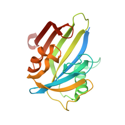
Crystal structure of zebrafish MTH1 in complex with TH588


Experimental Data Snapshot

  • Method: X-RAY DIFFRACTION
  • Resolution: 1.90 Å
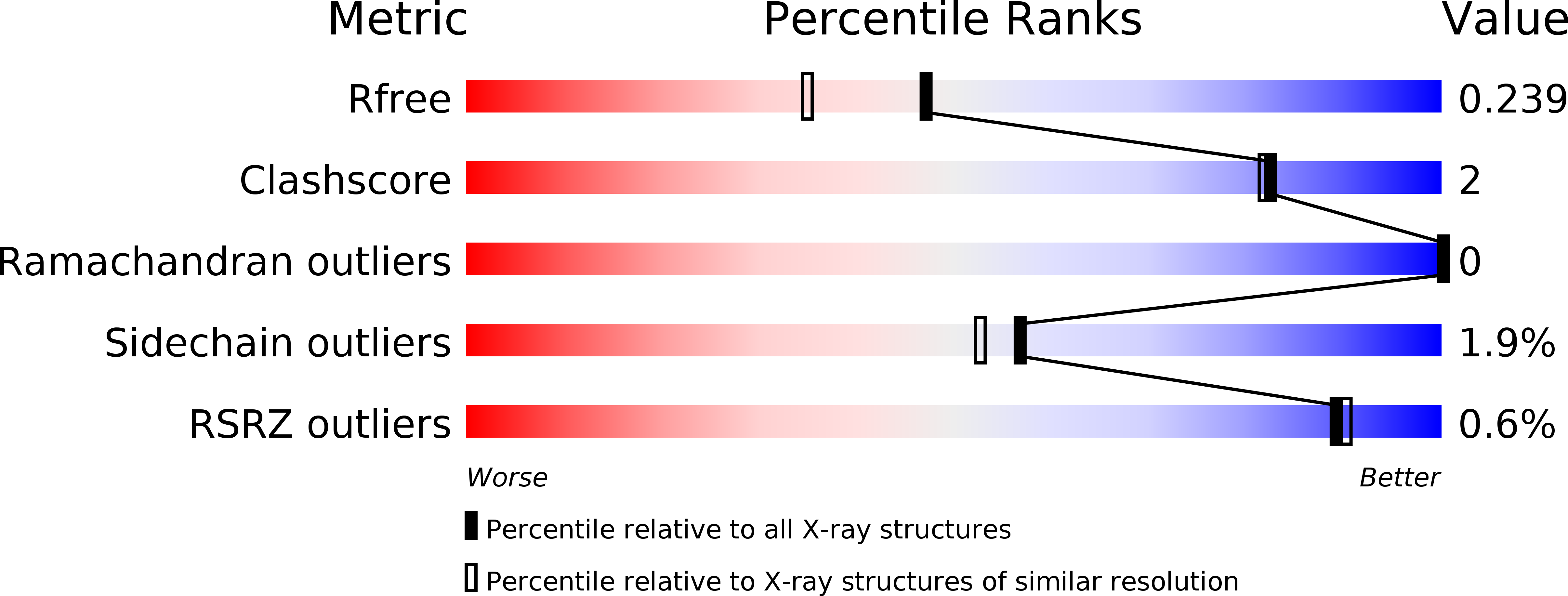
  • R-Value Free: 0.236 
  • R-Value Work: 0.184 
  • R-Value Observed: 0.186 

Starting Model: experimental
View more details

wwPDB Validation   3D Report Full Report


Ligand Structure Quality Assessment 


This is version 1.4 of the entry. See complete history


Literature

Hypoxic Signaling and the Cellular Redox Tumor Environment Determine Sensitivity to MTH1 Inhibition.

Brautigam, L.Pudelko, L.Jemth, A.S.Gad, H.Narwal, M.Gustafsson, R.Karsten, S.Carreras Puigvert, J.Homan, E.Berndt, C.Berglund, U.W.Stenmark, P.Helleday, T.

(2016) Cancer Res 76: 2366-2375

  • DOI: https://doi.org/10.1158/0008-5472.CAN-15-2380
  • Primary Citation of Related Structures:  
    5HZX

  • PubMed Abstract: 

    Cancer cells are commonly in a state of redox imbalance that drives their growth and survival. To compensate for oxidative stress induced by the tumor redox environment, cancer cells upregulate specific nononcogenic addiction enzymes, such as MTH1 (NUDT1), which detoxifies oxidized nucleotides. Here, we show that increasing oxidative stress in nonmalignant cells induced their sensitization to the effects of MTH1 inhibition, whereas decreasing oxidative pressure in cancer cells protected against inhibition. Furthermore, we purified zebrafish MTH1 and solved the crystal structure of MTH1 bound to its inhibitor, highlighting the zebrafish as a relevant tool to study MTH1 biology. Delivery of 8-oxo-dGTP and 2-OH-dATP to zebrafish embryos was highly toxic in the absence of MTH1 activity. Moreover, chemically or genetically mimicking activated hypoxia signaling in zebrafish revealed that pathologic upregulation of the HIF1α response, often observed in cancer and linked to poor prognosis, sensitized embryos to MTH1 inhibition. Using a transgenic zebrafish line, in which the cellular redox status can be monitored in vivo, we detected an increase in oxidative pressure upon activation of hypoxic signaling. Pretreatment with the antioxidant N-acetyl-L-cysteine protected embryos with activated hypoxia signaling against MTH1 inhibition, suggesting that the aberrant redox environment likely causes sensitization. In summary, MTH1 inhibition may offer a general approach to treat cancers characterized by deregulated hypoxia signaling or redox imbalance. Cancer Res; 76(8); 2366-75. ©2016 AACR.


  • Organizational Affiliation

    Science for Life Laboratory, Division of Translational Medicine and Chemical Biology, Department of Medical Biochemistry and Biophysics, Karolinska Institutet, Stockholm, Sweden. [email protected] [email protected].


Macromolecules
Find similar proteins by:  (by identity cutoff)  |  3D Structure
Entity ID: 1
MoleculeChains Sequence LengthOrganismDetailsImage
Nudix (Nucleoside diphosphate linked moiety X)-type motif 1
A, B
176Danio rerioMutation(s): 0 
Gene Names: nudt1
EC: 3.6.1 (UniProt), 3.6.1.56 (UniProt)
UniProt
Find proteins for Q7ZWC3 (Danio rerio)
Explore Q7ZWC3 
Go to UniProtKB:  Q7ZWC3
Entity Groups  
Sequence Clusters30% Identity50% Identity70% Identity90% Identity95% Identity100% Identity
UniProt GroupQ7ZWC3
Sequence Annotations
Expand
  • Reference Sequence
Small Molecules
Ligands 5 Unique
IDChains Name / Formula / InChI Key2D Diagram3D Interactions
2GE
Query on 2GE

Download Ideal Coordinates CCD File 
C [auth A],
I [auth B]
N~4~-cyclopropyl-6-(2,3-dichlorophenyl)pyrimidine-2,4-diamine
C13 H12 Cl2 N4
PNMYJIOQIAEYQL-UHFFFAOYSA-N
PEG
Query on PEG

Download Ideal Coordinates CCD File 
H [auth A]DI(HYDROXYETHYL)ETHER
C4 H10 O3
MTHSVFCYNBDYFN-UHFFFAOYSA-N
SO4
Query on SO4

Download Ideal Coordinates CCD File 
D [auth A],
E [auth A],
F [auth A],
J [auth B],
K [auth B]
SULFATE ION
O4 S
QAOWNCQODCNURD-UHFFFAOYSA-L
GOL
Query on GOL

Download Ideal Coordinates CCD File 
G [auth A]GLYCEROL
C3 H8 O3
PEDCQBHIVMGVHV-UHFFFAOYSA-N
ACT
Query on ACT

Download Ideal Coordinates CCD File 
L [auth B]ACETATE ION
C2 H3 O2
QTBSBXVTEAMEQO-UHFFFAOYSA-M
Experimental Data & Validation

Experimental Data

  • Method: X-RAY DIFFRACTION
  • Resolution: 1.90 Å
  • R-Value Free: 0.236 
  • R-Value Work: 0.184 
  • R-Value Observed: 0.186 
  • Space Group: P 21 21 2
Unit Cell:
Length ( Å )Angle ( ˚ )
a = 76.26α = 90
b = 71.202β = 90
c = 60.924γ = 90
Software Package:
Software NamePurpose
REFMACrefinement
XDSdata reduction
Aimlessdata scaling
MOLREPphasing

Structure Validation

View Full Validation Report


Ligand Structure Quality Assessment 


Entry History & Funding Information

Deposition Data


Funding OrganizationLocationGrant Number
Swedish Society for Medical researchSweden--
Karolinska Institute KID fundingSweden--
German Research FoundationGermany--
Knut and Alice Wallenberg FoundationSweden--
Swedish Foundation for Strategic ResearchSweden--
Swedish Cancer SocietySweden--
Swedish Research CouncilSweden--
Wenner-Gren FoundationSweden--
Goran Gustafsson FoundationSweden--
Swedish Children's Cancer FoundationSweden--
Swedish Pain Relief FoundationSweden--
Torsten and Ragnar Soderberg FoundationSweden--

Revision History  (Full details and data files)

  • Version 1.0: 2016-02-10
    Type: Initial release
  • Version 1.1: 2016-02-24
    Changes: Structure summary
  • Version 1.2: 2016-06-01
    Changes: Database references
  • Version 1.3: 2017-09-06
    Changes: Author supporting evidence
  • Version 1.4: 2024-01-10
    Changes: Data collection, Database references, Refinement description