5LAU

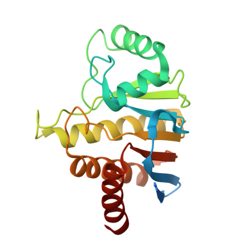
Oceanobacillus iheyensis macrodomain mutant G37V with ADPR


Experimental Data Snapshot

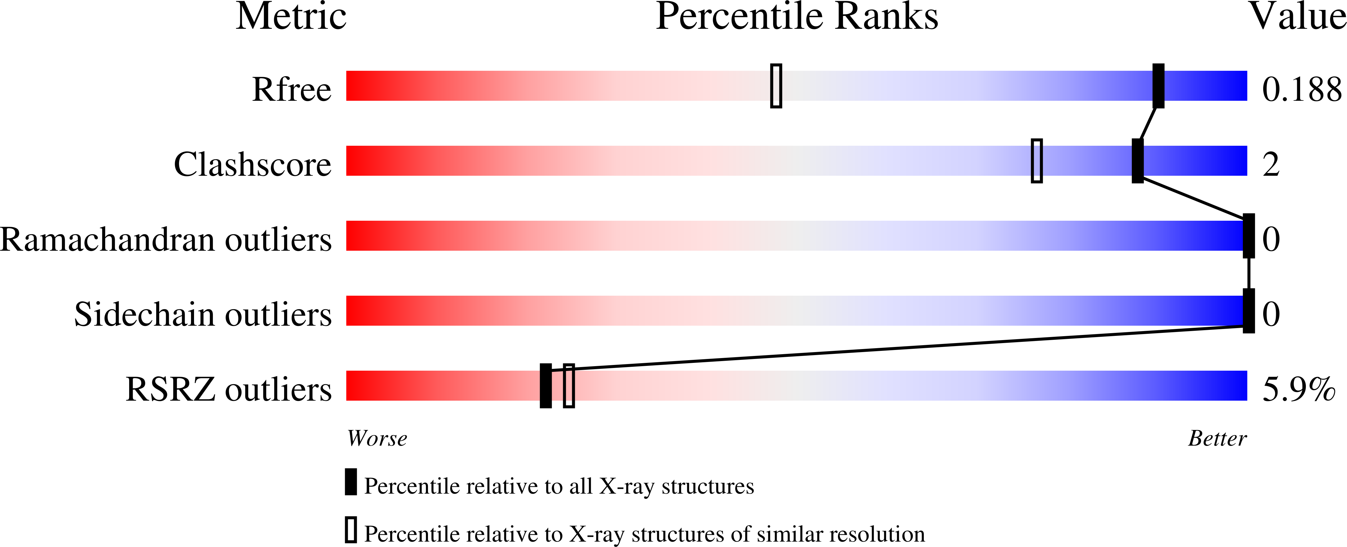
  • Method: X-RAY DIFFRACTION
  • Resolution: 1.35 Å
  • R-Value Free: 0.182 
  • R-Value Work: 0.154 
  • R-Value Observed: 0.156 

Starting Model: experimental
View more details

wwPDB Validation   3D Report Full Report


Ligand Structure Quality Assessment 


This is version 1.2 of the entry. See complete history


Literature

Structural and functional analysis of Oceanobacillus iheyensis macrodomain reveals a network of waters involved in substrate binding and catalysis.

Zapata-Perez, R.Gil-Ortiz, F.Martinez-Monino, A.B.Garcia-Saura, A.G.Juanhuix, J.Sanchez-Ferrer, A.

(2017) Open Biol 7

  • DOI: https://doi.org/10.1098/rsob.160327
  • Primary Citation of Related Structures:  
    5FUD, 5L9K, 5L9Q, 5LAU, 5LBP, 5LCC

  • PubMed Abstract: 

    Macrodomains are ubiquitous conserved domains that bind or transform ADP-ribose (ADPr) metabolites. In humans, they are involved in transcription, X-chromosome inactivation, neurodegeneration and modulating PARP1 signalling, making them potential targets for therapeutic agents. Unfortunately, some aspects related to the substrate binding and catalysis of MacroD-like macrodomains still remain unclear, since mutation of the proposed catalytic aspartate does not completely abolish enzyme activity. Here, we present a functional and structural characterization of a macrodomain from the extremely halotolerant and alkaliphilic bacterium Oceanobacillus iheyensis (OiMacroD), related to hMacroD1/hMacroD2, shedding light on substrate binding and catalysis. The crystal structures of D40A, N30A and G37V mutants, and those with MES, ADPr and ADP bound, allowed us to identify five fixed water molecules that play a significant role in substrate binding. Closure of the β6-α4 loop is revealed as essential not only for pyrophosphate recognition, but also for distal ribose orientation. In addition, a novel structural role for residue D40 is identified. Furthermore, it is revealed that OiMacroD not only catalyses the hydrolysis of O -acetyl-ADP-ribose but also reverses protein mono-ADP-ribosylation. Finally, mutant G37V supports the participation of a substrate-coordinated water molecule in catalysis that helps to select the proper substrate conformation.


  • Organizational Affiliation

    Department of Biochemistry and Molecular Biology-A, Faculty of Biology, Regional Campus of International Excellence 'Campus Mare Nostrum', University of Murcia, Campus Espinardo, 30100 Murcia, Spain.


Macromolecules
Find similar proteins by:  (by identity cutoff)  |  3D Structure
Entity ID: 1
MoleculeChains Sequence LengthOrganismDetailsImage
MacroD-type macrodomain208Oceanobacillus iheyensis HTE831Mutation(s): 1 
Gene Names: OB2288
UniProt
Find proteins for Q8EP31 (Oceanobacillus iheyensis (strain DSM 14371 / CIP 107618 / JCM 11309 / KCTC 3954 / HTE831))
Explore Q8EP31 
Go to UniProtKB:  Q8EP31
Entity Groups  
Sequence Clusters30% Identity50% Identity70% Identity90% Identity95% Identity100% Identity
UniProt GroupQ8EP31
Sequence Annotations
Expand
  • Reference Sequence
Small Molecules
Ligands 2 Unique
IDChains Name / Formula / InChI Key2D Diagram3D Interactions
AR6
Query on AR6

Download Ideal Coordinates CCD File 
B [auth A][(2R,3S,4R,5R)-5-(6-AMINOPURIN-9-YL)-3,4-DIHYDROXY-OXOLAN-2-YL]METHYL [HYDROXY-[[(2R,3S,4R,5S)-3,4,5-TRIHYDROXYOXOLAN-2-YL]METHOXY]PHOSPHORYL] HYDROGEN PHOSPHATE
C15 H23 N5 O14 P2
SRNWOUGRCWSEMX-ZQSHOCFMSA-N
GOL
Query on GOL

Download Ideal Coordinates CCD File 
C [auth A],
D [auth A]
GLYCEROL
C3 H8 O3
PEDCQBHIVMGVHV-UHFFFAOYSA-N
Experimental Data & Validation

Experimental Data

  • Method: X-RAY DIFFRACTION
  • Resolution: 1.35 Å
  • R-Value Free: 0.182 
  • R-Value Work: 0.154 
  • R-Value Observed: 0.156 
  • Space Group: I 2 3
Unit Cell:
Length ( Å )Angle ( ˚ )
a = 109.646α = 90
b = 109.646β = 90
c = 109.646γ = 90
Software Package:
Software NamePurpose
REFMACrefinement
XDSdata reduction
Aimlessdata scaling
PHASERphasing

Structure Validation

View Full Validation Report


Ligand Structure Quality Assessment 


Entry History & Funding Information

Deposition Data


Funding OrganizationLocationGrant Number
MINECO-FEDERSpainBIO2013-45336-R

Revision History  (Full details and data files)

  • Version 1.0: 2017-05-03
    Type: Initial release
  • Version 1.1: 2017-05-10
    Changes: Database references
  • Version 1.2: 2024-01-10
    Changes: Data collection, Database references, Refinement description, Structure summary